

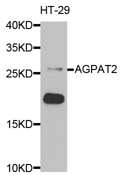

Anti-AGPAT2 Antibody
A29556
ApplicationsImmunoFluorescence, Western Blot, ImmunoHistoChemistry
Product group Antibodies
ReactivityHuman
Overview
- SupplierAntibodies.com
- Product NameAnti-AGPAT2 Antibody
- Delivery Days Customer7
- ApplicationsImmunoFluorescence, Western Blot, ImmunoHistoChemistry
- CertificationResearch Use Only
- ClonalityPolyclonal
- ConjugateUnconjugated
- Estimated Purity>95%
- HostRabbit
- Scientific DescriptionRabbit polyclonal antibody to AGPAT2
- ReactivityHuman
- UNSPSC12352203
Related products

Product group Antibodies
Anti-AGPAT2 Antibody Picoband(r)A04744-1-CARRIER-FREE
ApplicationsWestern Blot, ELISA, ImmunoHistoChemistry
ReactivityHuman, Mouse, Rat
TargetAGPAT2
- SizePrice
Product group Antibodies
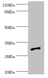
Anti-AGPAT2 Antibody144-66167
ApplicationsImmunoFluorescence, Western Blot
ReactivityHuman, Mouse
TargetAGPAT2
- SizePrice
Product group Antibodies
Agpat2 Polyclonal AntibodyBS-5032R
ApplicationsImmunoFluorescence, ELISA, ImmunoCytoChemistry, ImmunoHistoChemistry, ImmunoHistoChemistry Frozen, ImmunoHistoChemistry Paraffin
ReactivityBovine, Canine, Equine, Human, Mouse, Porcine, Rat
TargetAGPAT2
- SizePrice
Product group Antibodies
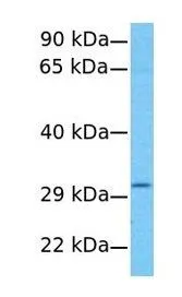
AGPAT2 AntibodyCSB-PA001450ESR2HU
ApplicationsWestern Blot, ELISA, ImmunoHistoChemistry
ReactivityHuman
TargetAGPAT2
- SizePrice
Product group Antibodies
Agpat2 Polyclonal AntibodyCAC10713
ApplicationsWestern Blot, ELISA, ImmunoHistoChemistry
TargetAGPAT2
- SizePrice
Product group Antibodies

AGPAT2 AntibodyLS-C402605
ApplicationsWestern Blot, ELISA
ReactivityHuman
TargetAGPAT2
- SizePrice
Product group Antibodies
AGPAT2 antibody, C-termGTX46672
ApplicationsWestern Blot
ReactivityHuman
TargetAGPAT2
- SizePrice

Product group Antibodies
Anti-AGPAT2 AntibodyHPA019544
ApplicationsWestern Blot, ImmunoHistoChemistry
ReactivityHuman
TargetAGPAT2
- SizePrice
Product group Antibodies
Anti-AGPAT2Y158051
ApplicationsELISA, ImmunoHistoChemistry
ReactivityHuman, Mouse, Rat
- SizePrice