


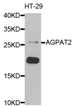
Figure 1. Western blot analysis of AGPAT2 using anti-AGPAT2 antibody (A04744-1). Electrophoresis was performed on a 5-20% SDS-PAGE gel at 70V (Stacking gel) / 90V (Resolving gel) for 2-3 hours. The sample well of each lane was loaded with 30 ug of sample under reducing conditions. Lane 1: human HEK293 whole cell lysates, Lane 2: human THP-1 whole cell lysates, Lane 3: rat liver tissue lysates, Lane 4: rat pancreas tissue lysates, Lane 5: mouse liver tissue lysates, Lane 6: mouse pancreas tissue lysates. After electrophoresis, proteins were transferred to a nitrocellulose membrane at 150 mA for 50-90 minutes. Blocked the membrane with 5% non-fat milk/TBS for 1.5 hour at RT. The membrane was incubated with rabbit anti-AGPAT2 antigen affinity purified polyclonal antibody (Catalog # A04744-1) at 0.5 microg/mL overnight at 4°C, then washed with TBS-0.1%Tween 3 times with 5 minutes each and probed with a goat anti-rabbit IgG-HRP secondary antibody at a dilution of 1:5000 for 1.5 hour at RT. The signal is developed using an Enhanced Chemiluminescent detection (ECL) kit (Catalog # EK1002) with Tanon 5200 system. A specific band was detected for AGPAT2 at approximately 33 kDa. The expected band size for AGPAT2 is at 33 kDa.
. AGPAT2 was detected in a paraffin-embedded section of human gall bladder adenosquamous carcinoma tissue. Heat mediated antigen retrieval was performed in EDTA buffer (pH 8.0, epitope retrieval solution). The tissue section was blocked with 10% goat serum. The tissue section was then incubated with 2 microg/ml rabbit anti-AGPAT2 Antibody (A04744-1) overnight at 4°C. Biotinylated goat anti-rabbit IgG was used as secondary antibody and incubated for 30 minutes at 37°C. The tissue section was developed using Strepavidin-Biotin-Complex (SABC) (Catalog # SA1022) with DAB as the chromogen.")
. AGPAT2 was detected in a paraffin-embedded section of mouse intestines tissue. Heat mediated antigen retrieval was performed in EDTA buffer (pH 8.0, epitope retrieval solution). The tissue section was blocked with 10% goat serum. The tissue section was then incubated with 2 microg/ml rabbit anti-AGPAT2 Antibody (A04744-1) overnight at 4°C. Biotinylated goat anti-rabbit IgG was used as secondary antibody and incubated for 30 minutes at 37°C. The tissue section was developed using Strepavidin-Biotin-Complex (SABC) (Catalog # SA1022) with DAB as the chromogen.")
. AGPAT2 was detected in a paraffin-embedded section of rat lung tissue. Heat mediated antigen retrieval was performed in EDTA buffer (pH 8.0, epitope retrieval solution). The tissue section was blocked with 10% goat serum. The tissue section was then incubated with 2 microg/ml rabbit anti-AGPAT2 Antibody (A04744-1) overnight at 4°C. Biotinylated goat anti-rabbit IgG was used as secondary antibody and incubated for 30 minutes at 37°C. The tissue section was developed using Strepavidin-Biotin-Complex (SABC) (Catalog # SA1022) with DAB as the chromogen.")
Figure 1. Western blot analysis of AGPAT2 using anti-AGPAT2 antibody (A04744-1). Electrophoresis was performed on a 5-20% SDS-PAGE gel at 70V (Stacking gel) / 90V (Resolving gel) for 2-3 hours. The sample well of each lane was loaded with 30 ug of sample under reducing conditions. Lane 1: human HEK293 whole cell lysates, Lane 2: human THP-1 whole cell lysates, Lane 3: rat liver tissue lysates, Lane 4: rat pancreas tissue lysates, Lane 5: mouse liver tissue lysates, Lane 6: mouse pancreas tissue lysates. After electrophoresis, proteins were transferred to a nitrocellulose membrane at 150 mA for 50-90 minutes. Blocked the membrane with 5% non-fat milk/TBS for 1.5 hour at RT. The membrane was incubated with rabbit anti-AGPAT2 antigen affinity purified polyclonal antibody (Catalog # A04744-1) at 0.5 microg/mL overnight at 4°C, then washed with TBS-0.1%Tween 3 times with 5 minutes each and probed with a goat anti-rabbit IgG-HRP secondary antibody at a dilution of 1:5000 for 1.5 hour at RT. The signal is developed using an Enhanced Chemiluminescent detection (ECL) kit (Catalog # EK1002) with Tanon 5200 system. A specific band was detected for AGPAT2 at approximately 33 kDa. The expected band size for AGPAT2 is at 33 kDa.
Anti-AGPAT2 Antibody Picoband(r)
A04744-1-CARRIER-FREE
ApplicationsWestern Blot, ELISA, ImmunoHistoChemistry
Product group Antibodies
ReactivityHuman, Mouse, Rat
TargetAGPAT2
Overview
- SupplierBoster Bio
- Product NameAnti-AGPAT2 Antibody Picoband(r)
- Delivery Days Customer9
- ApplicationsWestern Blot, ELISA, ImmunoHistoChemistry
- CertificationResearch Use Only
- ClonalityPolyclonal
- Concentration500 ug/ml
- Gene ID10555
- Target nameAGPAT2
- Target description1-acylglycerol-3-phosphate O-acyltransferase 2
- Target synonyms1-AGPAT2, BSCL, BSCL1, LPAAB, LPAAT-beta, LPLAT2, 1-acyl-sn-glycerol-3-phosphate acyltransferase beta, 1-AGP acyltransferase 2, 1-acylglycerol-3-phosphate O-acyltransferase 2 (lysophosphatidic acid acyltransferase, beta), lysophosphatidic acid acyltransferase-beta, lysophospholipid acyltransferase 2, testicular tissue protein Li 143
- HostRabbit
- IsotypeIgG
- Protein IDO15120
- Protein Name1-acyl-sn-glycerol-3-phosphate acyltransferase beta
- Scientific DescriptionBoster Bio Anti-AGPAT2 Antibody Picoband® catalog # A04744-1. Tested in ELISA, IHC, WB applications. This antibody reacts with Human, Mouse, Rat. The brand Picoband indicates this is a premium antibody that guarantees superior quality, high affinity, and strong signals with minimal background in Western blot applications. Only our best-performing antibodies are designated as Picoband, ensuring unmatched performance.
- ReactivityHuman, Mouse, Rat
- Storage Instruction-20°C,2°C to 8°C
- UNSPSC12352203
Related products
Product group Antibodies
Anti-AGPAT2 AntibodyA29556
ApplicationsImmunoFluorescence, Western Blot, ImmunoHistoChemistry
ReactivityHuman
- SizePrice
Product group Antibodies
Anti-AGPAT2 Antibody144-66167
ApplicationsImmunoFluorescence, Western Blot
ReactivityHuman, Mouse
TargetAGPAT2
- SizePrice
Product group Antibodies
Agpat2 Polyclonal AntibodyBS-5032R
ApplicationsImmunoFluorescence, ELISA, ImmunoCytoChemistry, ImmunoHistoChemistry, ImmunoHistoChemistry Frozen, ImmunoHistoChemistry Paraffin
ReactivityBovine, Canine, Equine, Human, Mouse, Porcine, Rat
TargetAGPAT2
- SizePrice
Product group Antibodies
AGPAT2 AntibodyCSB-PA001450ESR2HU
ApplicationsWestern Blot, ELISA, ImmunoHistoChemistry
ReactivityHuman
TargetAGPAT2
- SizePrice
Product group Antibodies
Agpat2 Polyclonal AntibodyCAC10713
ApplicationsWestern Blot, ELISA, ImmunoHistoChemistry
TargetAGPAT2
- SizePrice
Product group Antibodies

AGPAT2 AntibodyLS-C402605
ApplicationsWestern Blot, ELISA
ReactivityHuman
TargetAGPAT2
- SizePrice
Product group Antibodies
AGPAT2 antibody, C-termGTX46672
ApplicationsWestern Blot
ReactivityHuman
TargetAGPAT2
- SizePrice

Product group Antibodies
Anti-AGPAT2 AntibodyHPA019544
ApplicationsWestern Blot, ImmunoHistoChemistry
ReactivityHuman
TargetAGPAT2
- SizePrice
Product group Antibodies
Anti-AGPAT2Y158051
ApplicationsELISA, ImmunoHistoChemistry
ReactivityHuman, Mouse, Rat
- SizePrice