

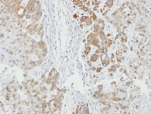
Immunohistochemical analysis of paraffin-embedded A549 xenograft, using AHA-1(GTX102321) antibody at 1:100 dilution.
Antigen Retrieval: Trilogy? (EDTA based, pH 8.0) buffer, 15min
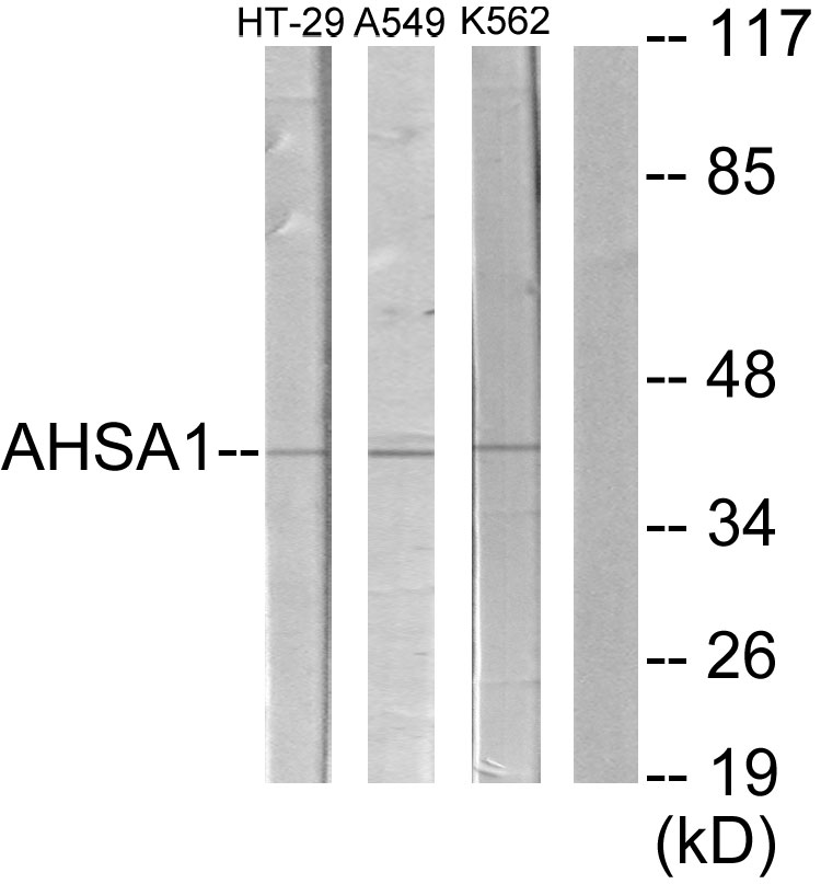
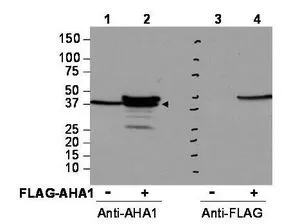
 A: JurKat 10% SDS PAGE AHA-1 antibody GTX102321 diluted at 1:1000")
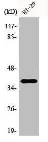
 antibody at 1:500 dilution.")
![AHA-1 antibody [N2C3] detects AHA-1 protein by immunofluorescent analysis. Sample: DIV9 rat E18 primary hippocampal neuron cells were fixed in 4% paraformaldehyde at RT for 15 min. Green: AHA-1 stained by AHA-1 antibody [N2C3] (GTX102321) diluted at 1:500. Red: beta Tubulin 3/ Tuj1, stained by beta Tubulin 3/ Tuj1 antibody [GT11710] (GTX631836) diluted at 1:500. Blue: Fluoroshield with DAPI (GTX30920).](https://www.genetex.com/upload/website/prouct_img/normal/GTX102321/GTX102321_40422_20181115_ICC_IF_R_w_23060100_618.webp "AHA-1 antibody [N2C3] detects AHA-1 protein by immunofluorescent analysis. Sample: DIV9 rat E18 primary hippocampal neuron cells were fixed in 4% paraformaldehyde at RT for 15 min. Green: AHA-1 stained by AHA-1 antibody [N2C3] (GTX102321) diluted at 1:500. Red: beta Tubulin 3/ Tuj1, stained by beta Tubulin 3/ Tuj1 antibody [GT11710] (GTX631836) diluted at 1:500. Blue: Fluoroshield with DAPI (GTX30920).")
![Wild-type (WT) and AHA-1 knockout (KO) HeLa cell extracts (30 μg) were separated by 10% SDS-PAGE, and the membrane was blotted with AHA-1 antibody [N2C3] (GTX102321) diluted at 1:30000. The HRP-conjugated anti-rabbit IgG antibody (GTX213110-01) was used to detect the primary antibody.](https://www.genetex.com/upload/website/prouct_img/normal/GTX102321/GTX102321_39673_20170601_WB_KO_watermark_w_23060100_758.webp "Wild-type (WT) and AHA-1 knockout (KO) HeLa cell extracts (30 μg) were separated by 10% SDS-PAGE, and the membrane was blotted with AHA-1 antibody [N2C3] (GTX102321) diluted at 1:30000. The HRP-conjugated anti-rabbit IgG antibody (GTX213110-01) was used to detect the primary antibody.")
Immunohistochemical analysis of paraffin-embedded A549 xenograft, using AHA-1(GTX102321) antibody at 1:100 dilution.
Antigen Retrieval: Trilogy? (EDTA based, pH 8.0) buffer, 15min
AHA-1 antibody [N2C3]
GTX102321
ApplicationsImmunoFluorescence, Western Blot, ImmunoCytoChemistry, ImmunoHistoChemistry, ImmunoHistoChemistry Paraffin
Product group Antibodies
ReactivityHuman, Rat
TargetAHSA1
Overview
- SupplierGeneTex
- Product NameAHA-1 antibody [N2C3]
- Delivery Days Customer9
- Application Supplier NoteWB: 1:500-1:30000. ICC/IF: 1:100-1:1000. IHC-P: 1:100-1:1000. *Optimal dilutions/concentrations should be determined by the researcher.Not tested in other applications.
- ApplicationsImmunoFluorescence, Western Blot, ImmunoCytoChemistry, ImmunoHistoChemistry, ImmunoHistoChemistry Paraffin
- CertificationResearch Use Only
- ClonalityPolyclonal
- Concentration1 mg/ml
- ConjugateUnconjugated
- Gene ID10598
- Target nameAHSA1
- Target descriptionactivator of HSP90 ATPase activity 1
- Target synonymsAHA1, C14orf3, hAha1, p38, activator of 90 kDa heat shock protein ATPase homolog 1, AHA1, activator of heat shock 90kDa protein ATPase homolog 1
- HostRabbit
- IsotypeIgG
- Protein IDO95433
- Protein NameActivator of 90 kDa heat shock protein ATPase homolog 1
- Scientific DescriptionCochaperone that stimulates HSP90 ATPase activity (By similarity). May affect a step in the endoplasmic reticulum to Golgi trafficking.
- ReactivityHuman, Rat
- Storage Instruction-20°C or -80°C,2°C to 8°C
- UNSPSC41116161
Datasheet
Related products
Product group Antibodies
Anti-AHSA1 (N-term) Antibody102-25481
ApplicationsFlow Cytometry, Western Blot
TargetAHSA1
- SizePrice

Product group Antibodies
Anti-AHA1/AHSA1 Antibody Picoband(r)A05733-2-CARRIER-FREE
ApplicationsFlow Cytometry, ImmunoFluorescence, Western Blot, ELISA, ImmunoCytoChemistry, ImmunoHistoChemistry
ReactivityHuman, Mouse, Rat
TargetAHSA1
- SizePrice
Product group Antibodies
Anti-AHSA1 AntibodyA101228
ApplicationsWestern Blot, ELISA
ReactivityHuman
- SizePrice
Product group Antibodies
AHSA1 Recombinant AntibodyBSM-62145R
ApplicationsImmunoPrecipitation, Western Blot
ReactivityHuman, Mouse, Rat
TargetAHSA1
- SizePrice
Product group Antibodies
AHSA1 AntibodyCSB-PA000833
ApplicationsWestern Blot, ELISA, ImmunoHistoChemistry
ReactivityHuman
TargetAHSA1
- SizePrice
Product group Antibodies
AHSA1 Polyclonal AntibodyCAC14079
ApplicationsWestern Blot, ELISA, ImmunoHistoChemistry
TargetAHSA1
- SizePrice
Product group Antibodies

AHSA1 / AHA1 AntibodyLS-C409561
ApplicationsWestern Blot
ReactivityHuman, Mouse, Rat
TargetAHSA1
- SizePrice
Product group Antibodies
AHA-1 antibody [N1C1]GTX102312
ApplicationsImmunoFluorescence, Western Blot, ImmunoCytoChemistry, ImmunoHistoChemistry, ImmunoHistoChemistry Paraffin
ReactivityHuman, Mouse
TargetAHSA1
- SizePrice

Product group Antibodies
Anti-AHSA1 AntibodyHPA000903
ApplicationsWestern Blot, ImmunoCytoChemistry, ImmunoHistoChemistry
ReactivityHuman
TargetAHSA1
- SizePrice
Product group Antibodies
AHA-1 antibodyGTX48723
ApplicationsImmunoPrecipitation, Western Blot, ELISA, ImmunoHistoChemistry
ReactivityHuman, Monkey
TargetAHSA1
- SizePrice