

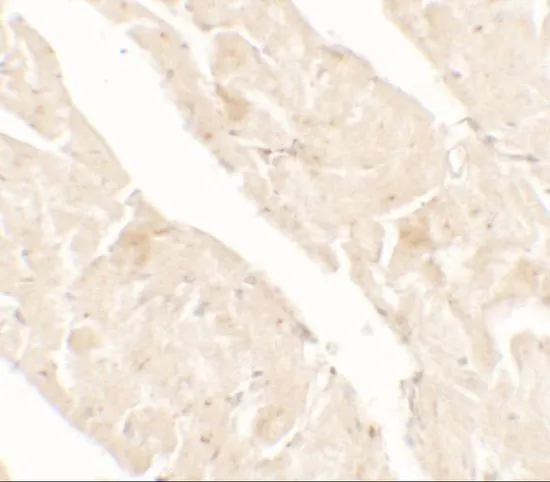
IHC-P analysis of rat small intestine tissue using GTX31896 AIMP2 antibody. Working concentration : 5 μg/ml

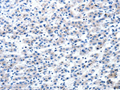
 the absence and (B) the presence of blocking peptide using GTX31896 AIMP2 antibody. Working concentration : 1 μg/ml")
IHC-P analysis of rat small intestine tissue using GTX31896 AIMP2 antibody. Working concentration : 5 μg/ml
AIMP2 antibody
GTX31896
ApplicationsWestern Blot, ELISA, ImmunoHistoChemistry, ImmunoHistoChemistry Paraffin
Product group Antibodies
ReactivityHuman, Rat
TargetAIMP2
Overview
- SupplierGeneTex
- Product NameAIMP2 antibody
- Delivery Days Customer9
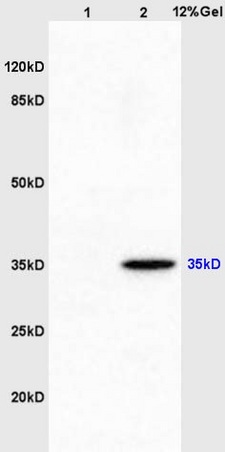
- Application Supplier NoteWB: 1 - 2 microg/mL. *Optimal dilutions/concentrations should be determined by the researcher.Not tested in other applications.
- ApplicationsWestern Blot, ELISA, ImmunoHistoChemistry, ImmunoHistoChemistry Paraffin
- CertificationResearch Use Only
- ClonalityPolyclonal
- Concentration1 mg/ml
- ConjugateUnconjugated
- Gene ID7965
- Target nameAIMP2
- Target descriptionaminoacyl tRNA synthetase complex interacting multifunctional protein 2
- Target synonymsHLD17, JTV-1, JTV1, P38, aminoacyl tRNA synthase complex-interacting multifunctional protein 2, ARS-interacting multi-functional protein 2, multisynthase complex auxiliary component p38, multisynthetase complex auxiliary component p38, protein JTV-1
- HostRabbit
- IsotypeIgG
- Protein IDQ13155
- Protein NameAminoacyl tRNA synthase complex-interacting multifunctional protein 2
- Scientific DescriptionThe JTV1 gene is located on chromosome 7p22 flanked by two genes, HRI and PMS2. JTV1 and HRI overlap slightly and are arranged in a tail-to-tail fashion. JTV1 and PMS2 are separated by approximately 200 base pairs and are arranged head-to-head. JTV1 is transcribed in the opposite direction compared to HRI and PMS2. The function of the JTV1 gene product is unknown. [provided by RefSeq, Jul 2008]
- ReactivityHuman, Rat
- Storage Instruction-20°C or -80°C,2°C to 8°C
- UNSPSC41116161
Datasheet
Related products
Product group Antibodies
Anti-JTV1 AntibodyA25007
ApplicationsWestern Blot
ReactivityHuman, Mouse, Rat
- SizePrice
Product group Antibodies
Anti-JTV1 Antibody101-10847
ApplicationsWestern Blot, ELISA
TargetAIMP2
- SizePrice
Product group Antibodies
References
AIMP2 Polyclonal AntibodyBS-2509R
ApplicationsImmunoFluorescence, Western Blot, ELISA, ImmunoCytoChemistry, ImmunoHistoChemistry, ImmunoHistoChemistry Frozen, ImmunoHistoChemistry Paraffin
ReactivityBovine, Human, Mouse, Porcine, Rabbit, Rat
TargetAIMP2
- SizePrice
Product group Antibodies
AIMP2 AntibodyCSB-PA151818
ApplicationsWestern Blot, ELISA, ImmunoHistoChemistry
ReactivityHuman, Mouse, Rat
TargetAIMP2
- SizePrice
![AIMP2 antibody [N2C3] detects AIMP2 protein at cytoplasm on human normal kidney by immunohistochemical analysis. Sample: Paraffin-embedded human normal kidney. AIMP2 antibody [N2C3] (GTX118650) diluted at 1:500.
Antigen Retrieval: Trilogy? (EDTA based, pH 8.0) buffer, 15min](https://www.genetex.com/upload/website/prouct_img/normal/GTX118650/GTX118650_40667_20141205_IHC_w_23060519_618.webp)
Product group Antibodies
AIMP2 antibody [N2C3]GTX118650
ApplicationsImmunoFluorescence, Western Blot, ImmunoCytoChemistry, ImmunoHistoChemistry, ImmunoHistoChemistry Paraffin
ReactivityHuman
TargetAIMP2
- SizePrice
![AIMP2 antibody [N1C1] detects AIMP2 protein at cytosol on mouse pancreas by immunohistochemical analysis. Sample: Paraffin-embedded mouse pancreas. AIMP2 antibody [N1C1] (GTX118947) dilution: 1:500.
Antigen Retrieval: Trilogy? (EDTA based, pH 8.0) buffer, 15min](https://www.genetex.com/upload/website/prouct_img/normal/GTX118947/GTX118947_40338_IHC_M_w_23060519_503.webp)
Product group Antibodies
AIMP2 antibody [N1C1]GTX118947
ApplicationsImmunoFluorescence, Western Blot, ImmunoCytoChemistry, ImmunoHistoChemistry, ImmunoHistoChemistry Paraffin
ReactivityHuman, Mouse
TargetAIMP2
- SizePrice

Product group Antibodies
Anti-AIMP2 AntibodyHPA019098
ApplicationsWestern Blot, ImmunoCytoChemistry, ImmunoHistoChemistry
ReactivityHuman, Mouse, Rat
TargetAIMP2
- SizePrice

Product group Antibodies
Anti-AIMP2/p38 Antibody Picoband(r)PA1481-CARRIER-FREE
ApplicationsWestern Blot, ImmunoHistoChemistry
ReactivityHuman, Mouse, Rat
TargetAIMP2
- SizePrice
Product group Antibodies

AIMP2 AntibodyLS-C763935
ApplicationsWestern Blot
ReactivityHuman, Rat
TargetAIMP2
- SizePrice