

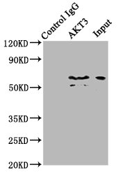
Immunoprecipitating AKT3 in mouse brain whole cell lysate Lane 1: Rabbit control IgG (1ug) instead of CSB-PA15929A0Rb in mouse brain whole cell lysate. For western blotting, a HRP-conjugated Protein G antibody was used as the secondary antibody (1/2000) Lane 2: CSB-PA15929A0Rb (6ug) + Mouse brain whole cell lysate (500ug) Lane 3: Mouse brain whole cell lysate (10ug)
Immunoprecipitating AKT3 in mouse brain whole cell lysate Lane 1: Rabbit control IgG (1ug) instead of CSB-PA15929A0Rb in mouse brain whole cell lysate. For western blotting, a HRP-conjugated Protein G antibody was used as the secondary antibody (1/2000) Lane 2: CSB-PA15929A0Rb (6ug) + Mouse brain whole cell lysate (500ug) Lane 3: Mouse brain whole cell lysate (10ug)
AKT3 Antibody
CSB-PA15929A0RB
ApplicationsImmunoPrecipitation, ELISA
Product group Antibodies
ReactivityHuman, Mouse
TargetAKT3
Overview
- SupplierCusabio
- Product NameAKT3 Antibody
- Delivery Days Customer20
- ApplicationsImmunoPrecipitation, ELISA
- CertificationResearch Use Only
- ClonalityPolyclonal
- ConjugateUnconjugated
- Gene ID10000
- Target nameAKT3
- Target descriptionAKT serine/threonine kinase 3
- Target synonymsMPPH, MPPH2, PKB-GAMMA, PKBG, PRKBG, RAC-PK-gamma, RAC-gamma, STK-2, RAC-gamma serine/threonine-protein kinase, PKB gamma, RAC-gamma serine/threonine protein kinase, v-akt murine thymoma viral oncogene homolog 3 (protein kinase B, gamma)
- HostRabbit
- IsotypeIgG
- Protein IDQ9Y243
- Protein NameRAC-gamma serine/threonine-protein kinase
- Scientific DescriptionAKT3 is one of 3 closely related serine/threonine-protein kinases (AKT1, AKT2 and AKT3) called the AKT kinase, and which regulate many processes including metabolism, proliferation, cell survival, growth and angiogenesis. This is mediated through serine and/or threonine phosphorylation of a range of downstream substrates. Over 100 substrate candidates have been reported so far, but for most of them, no isoform specificity has been reported. AKT3 is the least studied AKT isoform. It plays an important role in brain development and is crucial for the viability of malignant glioma cells. AKT3 isoform may also be the key molecule in up-regulation and down-regulation of MMP13 via IL13. Required for the coordination of mitochondrial biogenesis with growth factor-induced increases in cellular energy demands. Down-regulation by RNA interference reduces the expression of the phosphorylated form of BAD, resulting in the induction of caspase-dependent apoptosis.
- ReactivityHuman, Mouse
- Storage Instruction-20°C or -80°C
- UNSPSC41116161
Related products
Product group Antibodies
Anti-AKT3 AntibodyA83410
ApplicationsWestern Blot, ELISA, ImmunoHistoChemistry
ReactivityHuman, Mouse, Rat
- SizePrice
Product group Antibodies
Anti-AKT3 Antibody144-62553
ApplicationsWestern Blot, ImmunoHistoChemistry
ReactivityHuman, Mouse, Rat
TargetAKT3
- SizePrice
Product group Antibodies

AKT3 AntibodyLS-C747979
ApplicationsWestern Blot
ReactivityHuman, Mouse, Rat
TargetAKT3
- SizePrice

Product group Antibodies
Anti-AKT3 Antibody Picoband(r)A00520-2-CARRIER-FREE
ApplicationsWestern Blot, ELISA
ReactivityHuman, Mouse, Rat
TargetAKT3
- SizePrice
Product group Antibodies

Goat anti-AKT3EB06562
ApplicationsWestern Blot, ELISA, ImmunoHistoChemistry
ReactivityBovine, Canine, Human, Mouse, Rat
TargetAKT3
- SizePrice
Product group Antibodies
AKT3 Polyclonal AntibodyCAC15010
ApplicationsImmunoPrecipitation, ELISA
ReactivityMouse
TargetAKT3
- SizePrice

Product group Antibodies
Anti-AKT3 AntibodyHPA026441
ApplicationsWestern Blot, ImmunoHistoChemistry
ReactivityHuman
TargetAKT3
- SizePrice
Product group Antibodies
AKT3 antibodyGTX113312
ApplicationsImmunoFluorescence, Western Blot, ImmunoCytoChemistry, ImmunoHistoChemistry, ImmunoHistoChemistry Paraffin
ReactivityHuman, Mouse
TargetAKT3
- SizePrice