

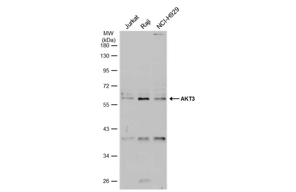
Various whole cell extracts (30 μg) were separated by 10% SDS-PAGE, and the membrane was blotted with AKT3 antibody (GTX113312) diluted at 1:1000. The HRP-conjugated anti-rabbit IgG antibody (GTX213110-01) was used to detect the primary antibody, and the signal was developed with Trident ECL plus-Enhanced.
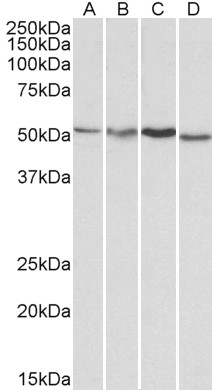
 A: Mouse brain 7.5% SDS PAGE GTX113312 diluted at 1:1000 The HRP-conjugated anti-rabbit IgG antibody (GTX213110-01) was used to detect the primary antibody.")
 diluted at 1:500. Antigen Retrieval: Citrate buffer, pH 6.0, 15 min")
 diluted at 1:500. Blue: Hoechst 33342 staining. Scale bar= 10 μm.")
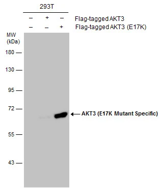
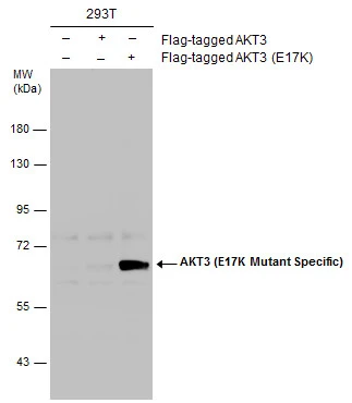
 and transfected (+) 293T whole cell extracts (30 μg) were separated by 10% SDS-PAGE, and the membrane was blotted with AKT3 antibody (GTX113312) diluted at 1:1000. The HRP-conjugated anti-rabbit IgG antibody (GTX213110-01) was used to detect the primary antibody.")
Various whole cell extracts (30 μg) were separated by 10% SDS-PAGE, and the membrane was blotted with AKT3 antibody (GTX113312) diluted at 1:1000. The HRP-conjugated anti-rabbit IgG antibody (GTX213110-01) was used to detect the primary antibody, and the signal was developed with Trident ECL plus-Enhanced.
AKT3 antibody
GTX113312
ApplicationsImmunoFluorescence, Western Blot, ImmunoCytoChemistry, ImmunoHistoChemistry, ImmunoHistoChemistry Paraffin
Product group Antibodies
ReactivityHuman, Mouse
TargetAKT3
Overview
- SupplierGeneTex
- Product NameAKT3 antibody
- Delivery Days Customer9
- Application Supplier NoteWB: 1:500-1:3000. ICC/IF: 1:100-1:1000. IHC-P: 1:100-1:1000. *Optimal dilutions/concentrations should be determined by the researcher.Not tested in other applications.
- ApplicationsImmunoFluorescence, Western Blot, ImmunoCytoChemistry, ImmunoHistoChemistry, ImmunoHistoChemistry Paraffin
- CertificationResearch Use Only
- ClonalityPolyclonal
- Concentration2.15 mg/ml
- ConjugateUnconjugated
- Gene ID10000
- Target nameAKT3
- Target descriptionAKT serine/threonine kinase 3
- Target synonymsMPPH, MPPH2, PKB-GAMMA, PKBG, PRKBG, RAC-PK-gamma, RAC-gamma, STK-2, RAC-gamma serine/threonine-protein kinase, PKB gamma, RAC-gamma serine/threonine protein kinase, v-akt murine thymoma viral oncogene homolog 3 (protein kinase B, gamma)
- HostRabbit
- IsotypeIgG
- Protein IDQ9Y243
- Protein NameRAC-gamma serine/threonine-protein kinase
- Scientific DescriptionThe protein encoded by this gene is a member of the AKT, also called PKB, serine/threonine protein kinase family. AKT kinases are known to be regulators of cell signaling in response to insulin and growth factors. They are involved in a wide variety of biological processes including cell proliferation, differentiation, apoptosis, tumorigenesis, as well as glycogen synthesis and glucose uptake. This kinase has been shown to be stimulated by platelet-derived growth factor (PDGF), insulin, and insulin-like growth factor 1 (IGF1). Alternatively splice transcript variants encoding distinct isoforms have been described. [provided by RefSeq]
- ReactivityHuman, Mouse
- Storage Instruction-20°C or -80°C,2°C to 8°C
- UNSPSC41116161
Datasheet
Related products
Product group Antibodies
Anti-AKT3 AntibodyA83410
ApplicationsWestern Blot, ELISA, ImmunoHistoChemistry
ReactivityHuman, Mouse, Rat
- SizePrice
Product group Antibodies
Anti-AKT3 Antibody144-62553
ApplicationsWestern Blot, ImmunoHistoChemistry
ReactivityHuman, Mouse, Rat
TargetAKT3
- SizePrice
Product group Antibodies

AKT3 AntibodyLS-C747979
ApplicationsWestern Blot
ReactivityHuman, Mouse, Rat
TargetAKT3
- SizePrice

Product group Antibodies
Anti-AKT3 Antibody Picoband(r)A00520-2-CARRIER-FREE
ApplicationsWestern Blot, ELISA
ReactivityHuman, Mouse, Rat
TargetAKT3
- SizePrice
Product group Antibodies
AKT3 AntibodyCSB-PA15929A0RB
ApplicationsImmunoPrecipitation, ELISA
ReactivityHuman, Mouse
TargetAKT3
- SizePrice
Product group Antibodies

Goat anti-AKT3EB06562
ApplicationsWestern Blot, ELISA, ImmunoHistoChemistry
ReactivityBovine, Canine, Human, Mouse, Rat
TargetAKT3
- SizePrice
Product group Antibodies
AKT3 Polyclonal AntibodyCAC15010
ApplicationsImmunoPrecipitation, ELISA
ReactivityMouse
TargetAKT3
- SizePrice
Product group Antibodies
AKT3 (E17K Mutant Specific) antibodyGTX132417
ApplicationsWestern Blot
ReactivityHuman
TargetAKT3
- SizePrice
Product group Antibodies
AKT3 (E17K Mutant Specific) antibodyGTX132418
ApplicationsWestern Blot
ReactivityHuman
TargetAKT3
- SizePrice