

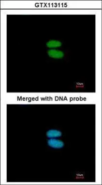
Immunofluorescence analysis of paraformaldehyde-fixed HeLa, using alpha Actinin 4(GTX113115) antibody at 1:200 dilution.
![alpha Actinin 4 antibody [N2C1], Internal detects alpha Actinin 4 protein by western blot analysis. A. 30 μg PC-12 whole cell lysate/extract 7.5% SDS-PAGE alpha Actinin 4 antibody [N2C1], Internal (GTX113115) dilution: 1:3000 The HRP-conjugated anti-rabbit IgG antibody (GTX213110-01) was used to detect the primary antibody.](https://www.genetex.com/upload/website/prouct_img/normal/GTX113115/GTX113115_40114_WB_R_w_23060500_829.webp "alpha Actinin 4 antibody [N2C1], Internal detects alpha Actinin 4 protein by western blot analysis. A. 30 μg PC-12 whole cell lysate/extract 7.5% SDS-PAGE alpha Actinin 4 antibody [N2C1], Internal (GTX113115) dilution: 1:3000 The HRP-conjugated anti-rabbit IgG antibody (GTX213110-01) was used to detect the primary antibody.")
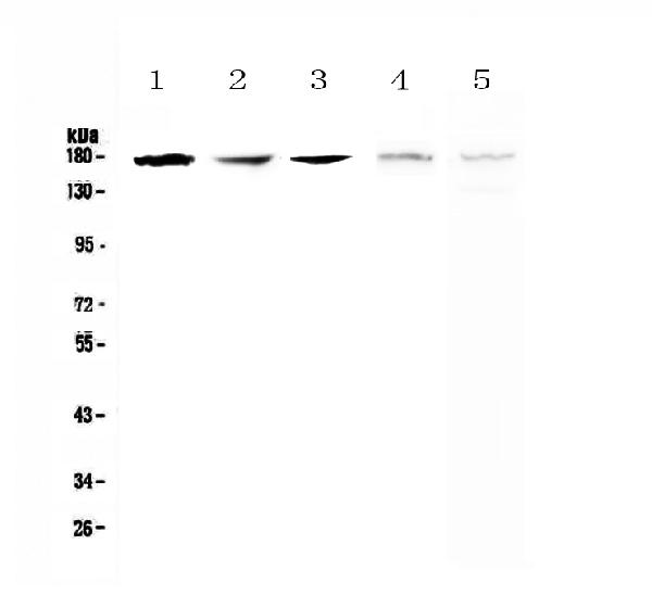
 A: Mouse brain 7.5% SDS PAGE GTX113115 diluted at 1:1000 The HRP-conjugated anti-rabbit IgG antibody (GTX213110-01) was used to detect the primary antibody.")
 antibody at 1:500 dilution.
Antigen Retrieval: Trilogy? (EDTA based, pH 8.0) buffer, 15min")
 A: NIH-3T3 7.5% SDS PAGE GTX113115 diluted at 1:1000 The HRP-conjugated anti-rabbit IgG antibody (GTX213110-01) was used to detect the primary antibody.")
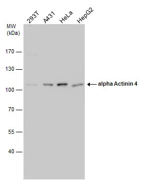
![Various whole cell extracts (30 μg) were separated by 7.5% SDS-PAGE, and the membrane was blotted with alpha Actinin 4 antibody [N2C1], Internal (GTX113115) diluted at 1:1000. The HRP-conjugated anti-rabbit IgG antibody (GTX213110-01) was used to detect the primary antibody.](https://www.genetex.com/upload/website/prouct_img/normal/GTX113115/GTX113115_44811_20220923_WB_24032600_886.webp "Various whole cell extracts (30 μg) were separated by 7.5% SDS-PAGE, and the membrane was blotted with alpha Actinin 4 antibody [N2C1], Internal (GTX113115) diluted at 1:1000. The HRP-conjugated anti-rabbit IgG antibody (GTX213110-01) was used to detect the primary antibody.")
Immunofluorescence analysis of paraformaldehyde-fixed HeLa, using alpha Actinin 4(GTX113115) antibody at 1:200 dilution.
alpha Actinin 4 antibody [N2C1], Internal
GTX113115
ApplicationsImmunoFluorescence, Western Blot, ImmunoCytoChemistry, ImmunoHistoChemistry, ImmunoHistoChemistry Paraffin
Product group Antibodies
ReactivityHuman, Mouse, Rat
TargetACTN4
Overview
- SupplierGeneTex
- Product Namealpha Actinin 4 antibody [N2C1], Internal
- Delivery Days Customer9
- Application Supplier NoteWB: 1:500-1:3000. ICC/IF: 1:100-1:1000. IHC-P: 1:100-1:1000. *Optimal dilutions/concentrations should be determined by the researcher.Not tested in other applications.
- ApplicationsImmunoFluorescence, Western Blot, ImmunoCytoChemistry, ImmunoHistoChemistry, ImmunoHistoChemistry Paraffin
- CertificationResearch Use Only
- ClonalityPolyclonal
- Concentration1.18 mg/ml
- ConjugateUnconjugated
- Gene ID81
- Target nameACTN4
- Target descriptionactinin alpha 4
- Target synonymsACTININ-4, FSGS, FSGS1, alpha-actinin-4, focal segmental glomerulosclerosis 1, non-muscle alpha-actinin 4
- HostRabbit
- IsotypeIgG
- Protein IDO43707
- Protein NameAlpha-actinin-4
- Scientific DescriptionAlpha actinins belong to the spectrin gene superfamily which represents a diverse group of cytoskeletal proteins, including the alpha and beta spectrins and dystrophins. Alpha actinin is an actin-binding protein with multiple roles in different cell types. In nonmuscle cells, the cytoskeletal isoform is found along microfilament bundles and adherens-type junctions, where it is involved in binding actin to the membrane. In contrast, skeletal, cardiac, and smooth muscle isoforms are localized to the Z-disc and analogous dense bodies, where they help anchor the myofibrillar actin filaments. This gene encodes a nonmuscle, alpha actinin isoform which is concentrated in the cytoplasm, and thought to be involved in metastatic processes. Mutations in this gene have been associated with focal and segmental glomerulosclerosis. [provided by RefSeq]
- ReactivityHuman, Mouse, Rat
- Storage Instruction-20°C or -80°C,2°C to 8°C
- UNSPSC12352203
References
- Grossi I, Radeghieri A, Paolini L, et al. MicroRNA‑34a‑5p expression in the plasma and in its extracellular vesicle fractions in subjects with Parkinson's disease: An exploratory study. Int J Mol Med. 2021,47(2):533-546. doi: 10.3892/ijmm.2020.4806Read this paper
Datasheet
Related products
Product group Antibodies

Anti-Alpha Actinin 4 [Ab01AA4]Ab02447-10.0
ApplicationsImmunoPrecipitation, ELISA, ImmunoHistoChemistry
ReactivityHuman
TargetACTN4
- SizePrice
Product group Antibodies
Anti-ACTN4 Antibody144-64273
ApplicationsImmunoFluorescence, Western Blot
ReactivityHuman, Mouse, Rat
TargetACTN4
- SizePrice
Product group Antibodies
Anti-IRE1/ERN1 Antibody Picoband(r)A00683-1-CARRIER-FREE
ApplicationsFlow Cytometry, ImmunoFluorescence, Western Blot, ELISA, ImmunoCytoChemistry
ReactivityHuman, Mouse, Rat
TargetACTN4
- SizePrice
![alpha Actinin 4 antibody [C2C3], C-term detects ACTN4 protein by western blot analysis. A. 30 μg Neuro2A whole cell lysate/extract B. 30 μg GL261 whole cell lysate/extract C. 30 μg C8D30 whole cell lysate/extract D. 30 μg NIH-3T3 whole cell lysate/extract E. 30 μg BCL-1 whole cell lysate/extract F. 30 μg Raw264.7 whole cell lysate/extract G. 30 μg C2C12 whole cell lysate/extract 7.5% SDS-PAGE alpha Actinin 4 antibody [C2C3], C-term (GTX101669) dilution: 1:1000 The HRP-conjugated anti-rabbit IgG antibody (GTX213110-01) was used to detect the primary antibody.](https://www.genetex.com/upload/website/prouct_img/normal/GTX101669/GTX101669_40205_WB_M_w_23060100_117.webp)
Product group Antibodies
References
ApplicationsImmunoFluorescence, Western Blot, ImmunoCytoChemistry, ImmunoHistoChemistry, ImmunoHistoChemistry Paraffin
ReactivityHuman, Mouse, Rat
TargetACTN4
- SizePrice
Product group Antibodies
ApplicationsImmunoFluorescence, Western Blot, ImmunoCytoChemistry
ReactivityHuman, Mouse, Rat
TargetACTN4
- SizePrice
![Various whole cell extracts (30 μg) were separated by 5% SDS-PAGE, and the membrane was blotted with alpha Actinin antibody [HL1692] (GTX637291) diluted at 1:20000. The HRP-conjugated anti-rabbit IgG antibody (GTX213110-01) was used to detect the primary antibody.](https://www.genetex.com/upload/website/prouct_img/normal/GTX637291/GTX637291_T-44767_20230210_WB_23021401_586.webp)
Product group Antibodies
alpha Actinin antibody [HL1692]GTX637291
ApplicationsImmunoFluorescence, Western Blot, ImmunoCytoChemistry, ImmunoHistoChemistry, ImmunoHistoChemistry Paraffin
ReactivityHuman, Mouse
TargetACTN4
- SizePrice
Product group Antibodies
Anti-ACTN4Y058985
ApplicationsWestern Blot, ELISA, ImmunoHistoChemistry
ReactivityHuman, Mouse, Rat
- SizePrice
Product group Antibodies
Actn4 Polyclonal AntibodyCAC10364
ApplicationsImmunoFluorescence, Western Blot, ELISA, ImmunoHistoChemistry
ReactivityRat
TargetACTN4
- SizePrice
Product group Antibodies
alpha Actinin 4 Recombinant AntibodyBSM-60423R
ApplicationsImmunoFluorescence, Western Blot, ImmunoCytoChemistry, ImmunoHistoChemistry, ImmunoHistoChemistry Frozen, ImmunoHistoChemistry Paraffin
ReactivityHuman, Mouse
TargetACTN4
- SizePrice