


Immunofluorescent staining of human cell line HEK 293 shows localization to nucleoli & cytosol.
Immunofluorescent staining of human cell line HEK 293 shows localization to nucleoli & cytosol.
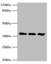
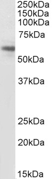
Anti-ACVR1 Antibody
HPA046514
ApplicationsImmunoCytoChemistry
Product group Antibodies
ReactivityHuman
TargetACVR1
Overview
- SupplierAtlas Antibodies
- Product NameAnti-ACVR1 Antibody
- Delivery Days Customer4
- ApplicationsImmunoCytoChemistry
- CertificationResearch Use Only
- ClonalityPolyclonal
- ConjugateUnconjugated
- Gene ID90
- Target nameACVR1
- Target descriptionactivin A receptor type 1
- Target synonymsACTRI, ACVR1A, ACVRLK2, ALK2, FOP, SKR1, TSRI, activin receptor type-1, TGF-B superfamily receptor type I, activin A receptor, type I, activin A receptor, type II-like kinase 2, activin receptor type I, activin receptor-like kinase 2, hydroxyalkyl-protein kinase, serine/threonine-protein kinase receptor R1
- HostRabbit
- IsotypeIgG
- Protein IDQ04771
- Protein NameActivin receptor type-1
- Scientific DescriptionRecombinant Protein Epitope Signature Tag (PrEST) antigen sequence
- ReactivityHuman
- Storage Instruction-20°C,2°C to 8°C
- UNSPSC41116161
Datasheet
MSDS
Related products
Product group Antibodies
ACVR1 AntibodyCSB-PA001257ESR2HU
ApplicationsImmunoFluorescence, Western Blot, ELISA, ImmunoHistoChemistry
ReactivityHuman, Mouse
TargetACVR1
- SizePrice
Product group Antibodies
ApplicationsImmunoPrecipitation, Western Blot, ImmunoCytoChemistry, ImmunoHistoChemistry
ReactivityMouse, Porcine, Rat
TargetACVR1
- SizePrice
Product group Antibodies
ApplicationsWestern Blot, ELISA
ReactivityHuman
- SizePrice

Product group Antibodies
References
ApplicationsImmunoPrecipitation, Western Blot
ReactivityHuman, Mouse, Rat
TargetACVR1
- SizePrice
Product group Antibodies
TargetACVR1
- SizePrice
Product group Antibodies

Goat anti-ACVR1, BiotinylatedEB08207-B
ApplicationsWestern Blot, ELISA, ImmunoHistoChemistry
ReactivityCanine, Human, Mouse, Rat
TargetACVR1
- SizePrice

Product group Antibodies
Anti-ACVR1 AntibodyHPA007505
ApplicationsImmunoHistoChemistry
ReactivityHuman
TargetACVR1
- SizePrice
Product group Antibodies
Anti-ACVR1 AntibodyHPA007505
ApplicationsImmunoHistoChemistry
ReactivityHuman
TargetACVR1
- SizePrice