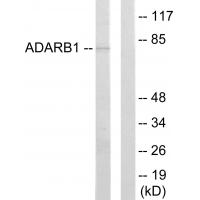
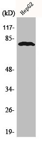
Bio-Connect

ADARB1 antibody

FNAB00151
Wuhan Fine Biotech
ApplicationsWestern Blot, ELISA
Product group Antibodies
TargetADARB1
Sign in to order and to see your custom pricing.
Large volume orders?
Order with a bulk request

Overview

  • Supplier
    Wuhan Fine Biotech
  • Product Name
    anti-ADARB1-antibody
  • Delivery Days Customer
    9
  • Applications
    Western Blot, ELISA
  • Certification
    Research Use Only
  • Clonality
    Polyclonal
  • Estimated Purity
    ≥95%
  • Gene ID104
  • Target name
    ADARB1
  • Target description
    adenosine deaminase RNA specific B1
  • Target synonyms
    ADAR2; adenosine deaminase, RNA-specific, B1 (homolog of rat RED1); double-stranded RNA-specific editase 1; DRABA2; DRADA2; dsRNA adenosine deaminase DRADA2; NEDHYMS; RED1; RNA editing deaminase 1; RNA-editing enzyme 1
  • Host
    Rabbit
  • Isotype
    IgG
  • Protein IDP78563
  • Protein Name
    Double-stranded RNA-specific editase 1
  • Storage Instruction
    -20°C
  • UNSPSC
    12352203

Related products

Western Blot analysis of HepG2 cells using ADAR2 Polyclonal Antibody
Product group Antibodies
Cusabio
ADARB1 AntibodyCSB-PA000821
ApplicationsWestern Blot, ELISA, ImmunoHistoChemistry
TargetADARB1
  • SizePrice
Product group Antibodies
References
GeneTex
ApplicationsImmunoFluorescence, Western Blot, ImmunoCytoChemistry
TargetADARB1
  • SizePrice
Immunohistochemical staining of human cerebral cortex shows strong nuclear positivity in neurons.
Product group Antibodies
Atlas Antibodies
ApplicationsImmunoHistoChemistry
TargetADARB1
  • SizePrice
Figure 1. Western blot analysis of RED1/ADARB1 using anti-RED1/ADARB1 antibody (A01810-2). Electrophoresis was performed on a 5-20% SDS-PAGE gel at 70V (Stacking gel) / 90V (Resolving gel) for 2-3 hours. The sample well of each lane was loaded with 30 ug of sample under reducing conditions. Lane 1: human SiHa whole cell lysates, Lane 2: human Hela whole cell lysates, Lane 3: human HEL whole cell lysates, Lane 4: rat brain tissue lysates, Lane 5: mouse brain tissue lysates. After electrophoresis, proteins were transferred to a nitrocellulose membrane at 150 mA for 50-90 minutes. Blocked the membrane with 5% non-fat milk/TBS for 1.5 hour at RT. The membrane was incubated with rabbit anti-RED1/ADARB1 antigen affinity purified polyclonal antibody (Catalog # A01810-2) at 0.5 microg/mL overnight at 4°C, then washed with TBS-0.1%Tween 3 times with 5 minutes each and probed with a goat anti-rabbit IgG-HRP secondary antibody at a dilution of 1:5000 for 1.5 hour at RT. The signal is developed using an Enhanced Chemiluminescent detection (ECL) kit (Catalog # EK1002) with Tanon 5200 system. A specific band was detected for RED1/ADARB1 at approximately 90 kDa. The expected band size for RED1/ADARB1 is at 81 kDa.
Product group Antibodies
Boster Bio
ApplicationsFlow Cytometry, Western Blot, ELISA
TargetADARB1
  • SizePrice
Product group Antibodies
RayBiotech
ApplicationsWestern Blot
TargetADARB1
  • SizePrice
Product group Antibodies
Biosynth
  • SizePrice
Product group Antibodies
LifeSpan BioSciences
ApplicationsWestern Blot, ELISA, ImmunoHistoChemistry
TargetADARB1
  • SizePrice
Anti-ADARB1 Antibody
Product group Antibodies
Antibodies.com
ApplicationsWestern Blot
  • SizePrice