


Immunohistochemical staining of human cerebral cortex shows strong nuclear positivity in neurons.
Immunohistochemical staining of human cerebral cortex shows strong nuclear positivity in neurons.
Anti-ADARB1 Antibody
HPA018277
ApplicationsImmunoHistoChemistry
Product group Antibodies
ReactivityHuman
TargetADARB1
Overview
- SupplierAtlas Antibodies
- Product NameAnti-ADARB1 Antibody
- Delivery Days Customer4
- ApplicationsImmunoHistoChemistry
- CertificationResearch Use Only
- ClonalityPolyclonal
- ConjugateUnconjugated
- Gene ID104
- Target nameADARB1
- Target descriptionadenosine deaminase RNA specific B1
- Target synonymsADAR2, DRABA2, DRADA2, NEDHYMS, RED1, double-stranded RNA-specific editase 1, RNA editing deaminase 1, RNA-editing enzyme 1, adenosine deaminase, RNA-specific, B1 (homolog of rat RED1), dsRNA adenosine deaminase DRADA2
- HostRabbit
- IsotypeIgG
- Protein IDP78563
- Protein NameDouble-stranded RNA-specific editase 1
- Scientific DescriptionRecombinant Protein Epitope Signature Tag (PrEST) antigen sequence
- ReactivityHuman
- Storage Instruction-20°C,2°C to 8°C
- UNSPSC41116161
Datasheet
MSDS
Related products
Product group Antibodies
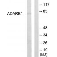
Anti-ADARB1 Antibody144-64636
ApplicationsWestern Blot
ReactivityHuman, Mouse, Rat
TargetADARB1
- SizePrice
Product group Antibodies
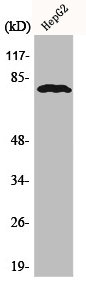
Anti-ADARB1 AntibodyA42124
ApplicationsWestern Blot
ReactivityHuman, Mouse, Rat
- SizePrice

Product group Antibodies
Anti-RED1/ADARB1 Antibody Picoband(r)A01810-2-CARRIER-FREE
ApplicationsFlow Cytometry, Western Blot, ELISA
ReactivityHuman, Mouse, Rat
TargetADARB1
- SizePrice
Product group Antibodies
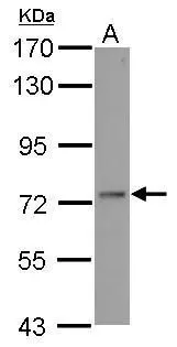
ADARB1 AntibodyCSB-PA000821
ApplicationsWestern Blot, ELISA, ImmunoHistoChemistry
ReactivityHuman, Mouse, Rat
TargetADARB1
- SizePrice
Product group Antibodies

ApplicationsWestern Blot, ELISA, ImmunoHistoChemistry
ReactivityHuman, Mouse, Rat
TargetADARB1
- SizePrice
Product group Antibodies
ADAR2 antibodyGTX114237
ApplicationsImmunoFluorescence, Western Blot, ImmunoCytoChemistry
ReactivityHuman
TargetADARB1
- SizePrice