


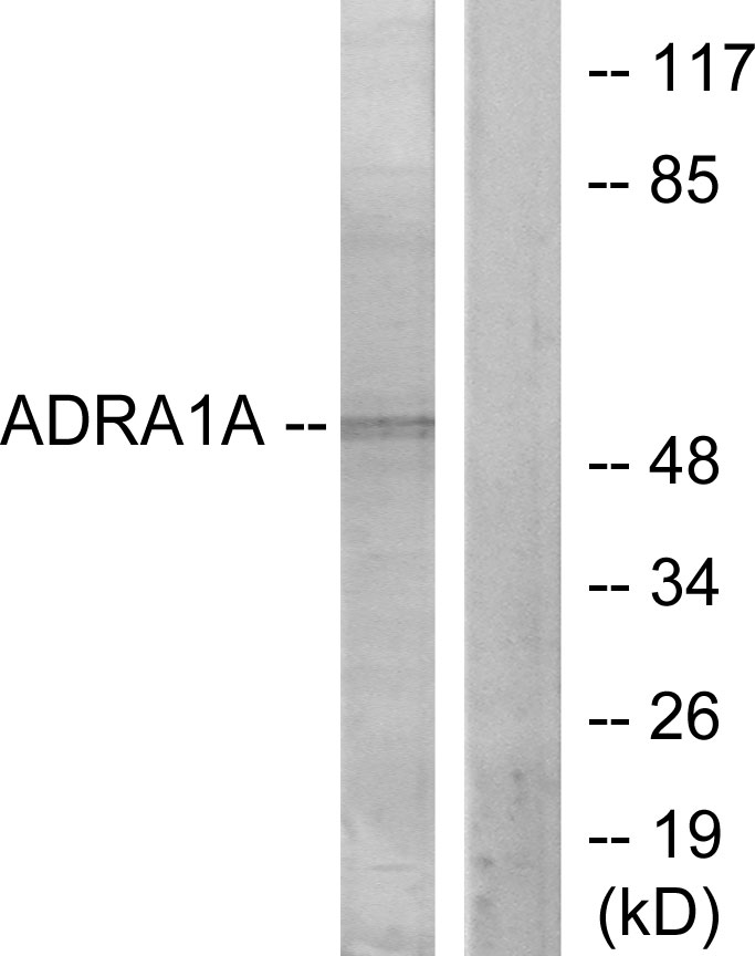
Figure 1. Western blot analysis of ADRA1A using anti-ADRA1A antibody (PB9752). Electrophoresis was performed on a 5-20% SDS-PAGE gel at 70V (Stacking gel) / 90V (Resolving gel) for 2-3 hours. Lane 1: Rat Cardiac Muscle Tissue Lysate at 50ug, Lane 2: Rat Brain Tissue Lysate at 50ug, Lane 3: Rat Liver Tissue Lysate at 50ug, Lane 4: Mouse Liver Tissue Lysate at 50ug, Lane 5: Mouse Lung Tissue Lysate at 50ug, Lane 6: 22RV1 Whole Cell Lysate at 40ug, Lane 7: SMMC Whole Cell Lysate at 40ug. After electrophoresis, proteins were transferred to a nitrocellulose membrane at 150 mA for 50-90 minutes. Blocked the membrane with 5% non-fat milk/TBS for 1.5 hour at RT. The membrane was incubated with rabbit anti-ADRA1A antigen affinity purified polyclonal antibody (Catalog # PB9752) at 0.5 microg/mL overnight at 4°C, then washed with TBS-0.1%Tween 3 times with 5 minutes each and probed with a goat anti-rabbit IgG-HRP secondary antibody at a dilution of 1:5000 for 1.5 hour at RT. The signal is developed using an Enhanced Chemiluminescent detection (ECL) kit (Catalog # EK1002) with Tanon 5200 system. A specific band was detected for ADRA1A at approximately 51 kDa. The expected band size for ADRA1A is at 51 kDa.
. ADRA1A was detected in paraffin-embedded section of human liver cancer tissues. Heat mediated antigen retrieval was performed in citrate buffer (pH6, epitope retrieval solution) for 20 mins. The tissue section was blocked with 10% goat serum. The tissue section was then incubated with 1microg/ml rabbit anti-ADRA1A Antibody (PB9752) overnight at 4°C. Biotinylated goat anti-rabbit IgG was used as secondary antibody and incubated for 30 minutes at 37°C. The tissue section was developed using Strepavidin-Biotin-Complex (SABC)(Catalog # SA1022) with DAB as the chromogen.")
. ADRA1A was detected in paraffin-embedded section of rat brain tissue tissues. Heat mediated antigen retrieval was performed in citrate buffer (pH6, epitope retrieval solution) for 20 mins. The tissue section was blocked with 10% goat serum. The tissue section was then incubated with 1microg/ml rabbit anti-ADRA1A Antibody (PB9752) overnight at 4°C. Biotinylated goat anti-rabbit IgG was used as secondary antibody and incubated for 30 minutes at 37°C. The tissue section was developed using Strepavidin-Biotin-Complex (SABC)(Catalog # SA1022) with DAB as the chromogen.")
. ADRA1A was detected in paraffin-embedded section of rat brain tissue tissues. Heat mediated antigen retrieval was performed in citrate buffer (pH6, epitope retrieval solution) for 20 mins. The tissue section was blocked with 10% goat serum. The tissue section was then incubated with 1microg/ml rabbit anti-ADRA1A Antibody (PB9752) overnight at 4°C. Biotinylated goat anti-rabbit IgG was used as secondary antibody and incubated for 30 minutes at 37°C. The tissue section was developed using Strepavidin-Biotin-Complex (SABC)(Catalog # SA1022) with DAB as the chromogen.")
. Overlay histogram showing A431 cells stained with PB9752 (Blue line).The cells were blocked with 10% normal goat serum. And then incubated with rabbit anti-ADRA1A Antibody (PB9752,1microg/1x106 cells) for 30 min at 20°C. DyLight®488 conjugated goat anti-rabbit IgG (BA1127, 5-10microg/1x106 cells) was used as secondary antibody for 30 minutes at 20°C. Isotype control antibody (Green line) was rabbit IgG (1microg/1x106) used under the same conditions. Unlabelled sample (Red line) was also used as a control.")
Figure 1. Western blot analysis of ADRA1A using anti-ADRA1A antibody (PB9752). Electrophoresis was performed on a 5-20% SDS-PAGE gel at 70V (Stacking gel) / 90V (Resolving gel) for 2-3 hours. Lane 1: Rat Cardiac Muscle Tissue Lysate at 50ug, Lane 2: Rat Brain Tissue Lysate at 50ug, Lane 3: Rat Liver Tissue Lysate at 50ug, Lane 4: Mouse Liver Tissue Lysate at 50ug, Lane 5: Mouse Lung Tissue Lysate at 50ug, Lane 6: 22RV1 Whole Cell Lysate at 40ug, Lane 7: SMMC Whole Cell Lysate at 40ug. After electrophoresis, proteins were transferred to a nitrocellulose membrane at 150 mA for 50-90 minutes. Blocked the membrane with 5% non-fat milk/TBS for 1.5 hour at RT. The membrane was incubated with rabbit anti-ADRA1A antigen affinity purified polyclonal antibody (Catalog # PB9752) at 0.5 microg/mL overnight at 4°C, then washed with TBS-0.1%Tween 3 times with 5 minutes each and probed with a goat anti-rabbit IgG-HRP secondary antibody at a dilution of 1:5000 for 1.5 hour at RT. The signal is developed using an Enhanced Chemiluminescent detection (ECL) kit (Catalog # EK1002) with Tanon 5200 system. A specific band was detected for ADRA1A at approximately 51 kDa. The expected band size for ADRA1A is at 51 kDa.
Anti-alpha 1d Adrenergic Receptor/ADRA1A Antibody Picoband(r)
PB9752-CARRIER-FREE
ApplicationsWestern Blot
Product group Antibodies
ReactivityHuman, Mouse, Rat
TargetADRA1A
Overview
- SupplierBoster Bio
- Product NameAnti-alpha 1d Adrenergic Receptor/ADRA1A Antibody Picoband(r)
- Delivery Days Customer9
- Application Supplier NoteTested Species: In-house tested species with positive results. Other applications have not been tested. Optimal dilutions should be determined by end users.
- ApplicationsWestern Blot
- CertificationResearch Use Only
- ClonalityPolyclonal
- Concentration500 ug/ml
- Gene ID148
- Target nameADRA1A
- Target descriptionadrenoceptor alpha 1A
- Target synonymsADRA1C, ADRA1L1, ALPHA1AAR, alpha-1A adrenergic receptor, G protein coupled receptor, alpha-1A adrenoceptor, alpha-1A adrenoreceptor, alpha-1C adrenergic receptor
- HostRabbit
- IsotypeIgG
- Protein IDP35348
- Protein NameAlpha-1A adrenergic receptor
- Scientific DescriptionBoster Bio Anti-alpha 1d Adrenergic Receptor/ADRA1A Antibody Picoband® catalog # PB9752. Tested in WB applications. This antibody reacts with Human, Mouse, Rat. The brand Picoband indicates this is a premium antibody that guarantees superior quality, high affinity, and strong signals with minimal background in Western blot applications. Only our best-performing antibodies are designated as Picoband, ensuring unmatched performance.
- ReactivityHuman, Mouse, Rat
- Storage Instruction-20°C,2°C to 8°C
- UNSPSC12352203
Related products
Product group Antibodies
ADRA1A AntibodyCSB-PA001385PA01HU
ApplicationsImmunoFluorescence, Western Blot, ELISA
ReactivityHuman
TargetADRA1A
- SizePrice
Product group Antibodies
Anti-ADRA1A AntibodyA100623
ApplicationsWestern Blot, ELISA
ReactivityHuman
- SizePrice
Product group Antibodies

Goat anti-ADRA1AEB11923
ApplicationsWestern Blot, ELISA
ReactivityHuman
TargetADRA1A
- SizePrice

Product group Antibodies
Anti-ADRA1A AntibodyHPA076693
ApplicationsWestern Blot, ImmunoCytoChemistry
ReactivityHuman
TargetADRA1A
- SizePrice
Product group Antibodies

ADRA1A Antibody (Internal)LS-C368567
ApplicationsImmunoFluorescence, Western Blot, ImmunoCytoChemistry
ReactivityBovine, Canine, Human, Rat
TargetADRA1A
- SizePrice
Product group Antibodies
ADRA1A Polyclonal AntibodyCAC15719
ApplicationsImmunoFluorescence, Western Blot, ELISA
TargetADRA1A
- SizePrice
![alpha 1a Adrenergic Receptor antibody [HL3291] detects alpha 1a Adrenergic Receptor protein by immunofluorescent analysis. Sample: 22RV1 cells were fixed in ice-cold MeOH for 5 min. Green: alpha 1a Adrenergic Receptor stained by alpha 1a Adrenergic Receptor antibody [HL3291] (GTX640945) diluted at 1:500. Blue: Fluoroshield with DAPI (GTX30920).](https://www.genetex.com/upload/website/prouct_img/normal/GTX640945/GTX640945_T-45530_20240927_ICC_IF_24111918_905.webp)
Product group Antibodies
ApplicationsImmunoFluorescence, Western Blot, ImmunoCytoChemistry
ReactivityHuman
TargetADRA1A
- SizePrice
Product group Antibodies
References
ADRA1A Polyclonal AntibodyBS-0600R
ApplicationsFlow Cytometry, ImmunoFluorescence, Western Blot, ELISA, ImmunoCytoChemistry, ImmunoHistoChemistry, ImmunoHistoChemistry Frozen, ImmunoHistoChemistry Paraffin
ReactivityBovine, Canine, Chicken, Equine, Guinea Pig, Human, Mouse, Porcine, Rabbit, Rat, Sheep
TargetADRA1A
- SizePrice