


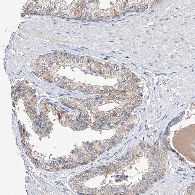
Immunohistochemical staining of human placenta shows distinct nuclear positivity in trophoblastic cells.

Immunohistochemical staining of human placenta shows distinct nuclear positivity in trophoblastic cells.
Anti-AMD1 Antibody
HPA029281
ApplicationsImmunoCytoChemistry, ImmunoHistoChemistry
Product group Antibodies
ReactivityHuman
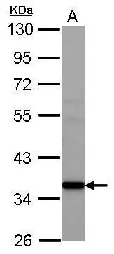
TargetAMD1
Overview
- SupplierAtlas Antibodies
- Product NameAnti-AMD1 Antibody
- Delivery Days Customer4
- ApplicationsImmunoCytoChemistry, ImmunoHistoChemistry
- CertificationResearch Use Only
- ClonalityPolyclonal
- ConjugateUnconjugated
- Gene ID262
- Target nameAMD1
- Target descriptionadenosylmethionine decarboxylase 1
- Target synonymsADOMETDC, AMD, SAMDC, S-adenosylmethionine decarboxylase proenzyme, S-adenosylmethionine decarboxylase 1
- HostRabbit
- IsotypeIgG
- Protein IDP17707
- Protein NameS-adenosylmethionine decarboxylase proenzyme
- Scientific DescriptionRecombinant Protein Epitope Signature Tag (PrEST) antigen sequence
- ReactivityHuman
- Storage Instruction-20°C,2°C to 8°C
- UNSPSC41116161
Datasheet
MSDS
Related products
Product group Antibodies
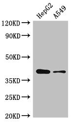
AMD1 AntibodyCSB-PA001568LA01HU
ApplicationsImmunoFluorescence, Western Blot, ELISA
ReactivityHuman
TargetAMD1
- SizePrice

Product group Antibodies
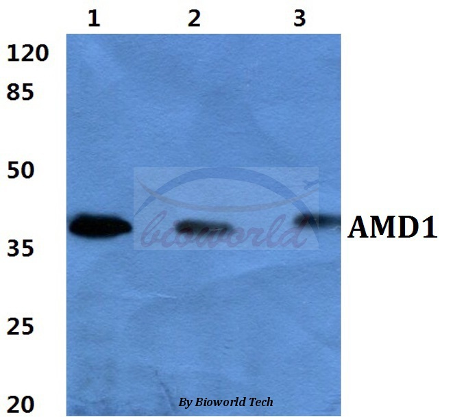
Anti-AMD1 Antibody Picoband(r)A06146-CARRIER-FREE
ApplicationsWestern Blot, ImmunoHistoChemistry
ReactivityHuman, Mouse, Rat
TargetAMD1
- SizePrice
Product group Antibodies
Anti-AMD1 AntibodyA28593
ApplicationsWestern Blot
ReactivityHuman, Mouse, Rat
- SizePrice
Product group Antibodies
Anti-AMD1 AntibodyHPA029282
ApplicationsImmunoHistoChemistry
ReactivityHuman
TargetAMD1
- SizePrice
Product group Antibodies

AMD / AMD1 AntibodyLS-C402769
ApplicationsELISA, ImmunoHistoChemistry
ReactivityHuman, Mouse, Rat
TargetAMD1
- SizePrice
Product group Antibodies
AMD1/SAMDC Polyclonal AntibodyBS-1887R
ApplicationsImmunoFluorescence, ELISA, ImmunoCytoChemistry, ImmunoHistoChemistry, ImmunoHistoChemistry Frozen, ImmunoHistoChemistry Paraffin
ReactivityBovine, Canine, Equine, Human, Mouse, Porcine, Rabbit, Rat
TargetAMD1
- SizePrice
Product group Antibodies
AMD1 antibodyGTX119689
ApplicationsWestern Blot
ReactivityHuman
TargetAMD1
- SizePrice