

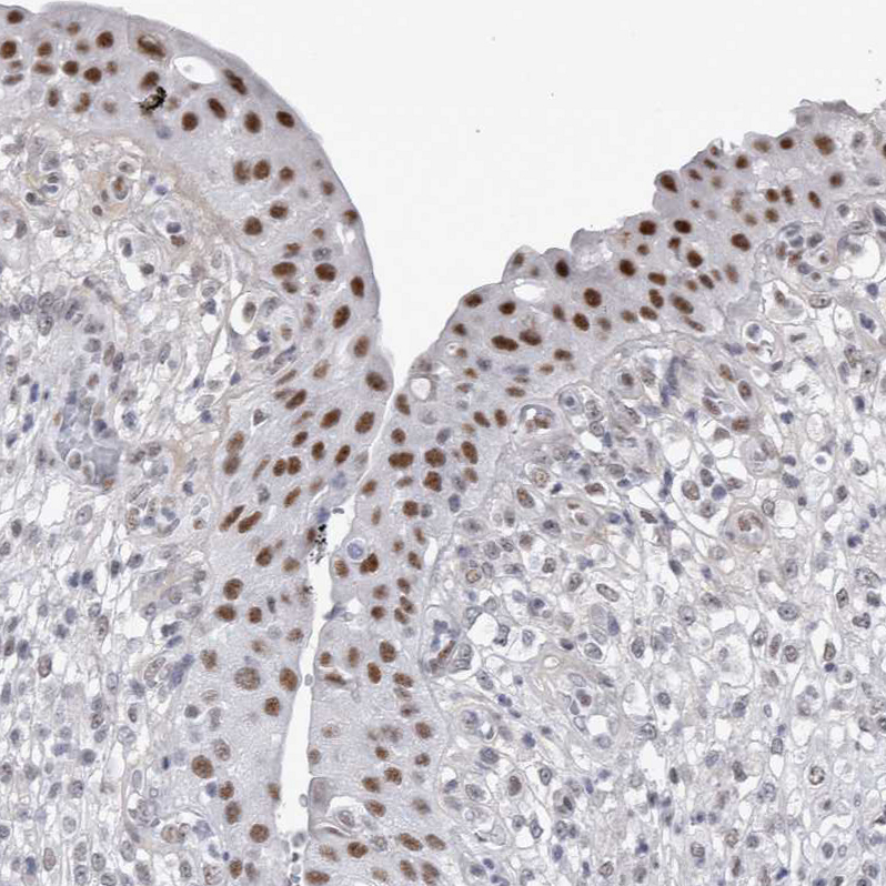
Immunohistochemical staining of human urinary bladder shows strong nuclear positivity in urothelial cells.

Immunohistochemical staining of human urinary bladder shows strong nuclear positivity in urothelial cells.
Anti-API5 Antibody
HPA026598
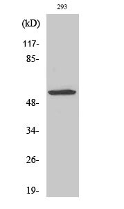
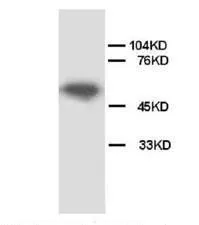
ApplicationsWestern Blot, ImmunoCytoChemistry, ImmunoHistoChemistry
Product group Antibodies
ReactivityHuman, Mouse, Rat
TargetAPI5
Overview
- SupplierAtlas Antibodies
- Product NameAnti-API5 Antibody
- Delivery Days Customer4
- ApplicationsWestern Blot, ImmunoCytoChemistry, ImmunoHistoChemistry
- CertificationResearch Use Only
- ClonalityPolyclonal
- ConjugateUnconjugated
- Gene ID8539
- Target nameAPI5
- Target descriptionapoptosis inhibitor 5
- Target synonymsAAC-11, AAC11, apoptosis inhibitor 5, FIF, antiapoptosis clone 11 protein, cell migration-inducing gene 8 protein, fibroblast growth factor 2-interacting factor 2, migration-inducing protein MIG8
- HostRabbit
- IsotypeIgG
- Protein IDQ9BZZ5
- Protein NameApoptosis inhibitor 5
- Scientific DescriptionRecombinant Protein Epitope Signature Tag (PrEST) antigen sequence
- ReactivityHuman, Mouse, Rat
- Storage Instruction-20°C,2°C to 8°C
- UNSPSC41116161
Datasheet
MSDS
Related products

Product group Antibodies
API5 AntibodyCSB-PA000902
ApplicationsWestern Blot, ELISA, ImmunoHistoChemistry
ReactivityHuman, Mouse
TargetAPI5
- SizePrice
Product group Antibodies
Anti-API5 AntibodyA35992
ApplicationsWestern Blot, ELISA, ImmunoHistoChemistry
ReactivityHuman, Mouse
- SizePrice
Product group Antibodies

API5 AntibodyLS-C406134
ApplicationsWestern Blot, ELISA
ReactivityHuman
TargetAPI5
- SizePrice
Product group Antibodies
API5 Polyclonal AntibodyCAC15024
ApplicationsImmunoFluorescence, Western Blot, ELISA
ReactivityMouse
TargetAPI5
- SizePrice

Product group Antibodies
Anti-Apoptosis inhibitor 5/API5 Antibody Picoband(r)PA1009-CARRIER-FREE
ApplicationsImmunoFluorescence, Western Blot, ImmunoCytoChemistry, ImmunoHistoChemistry, ImmunoHistoChemistry Frozen
ReactivityHamster, Human, Mouse, Rat
TargetAPI5
- SizePrice
Product group Antibodies
API5 antibodyGTX38554
ApplicationsWestern Blot, ImmunoHistoChemistry, ImmunoHistoChemistry Paraffin
ReactivityHuman, Rat
TargetAPI5
- SizePrice
Product group Antibodies
API5 Polyclonal AntibodyBS-1252R
ApplicationsImmunoFluorescence, Western Blot, ImmunoHistoChemistry, ImmunoHistoChemistry Frozen, ImmunoHistoChemistry Paraffin
ReactivityBovine, Canine, Equine, Guinea Pig, Human, Mouse, Porcine, Rat
TargetAPI5
- SizePrice
Product group Antibodies
Anti-API5 (C-term) Antibody102-23357
ApplicationsWestern Blot
TargetAPI5
- SizePrice