


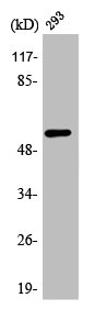
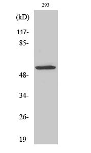
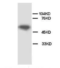
Figure 1. Western blot analysis of API5 using anti-API5 antibody (PA1009). Electrophoresis was performed on a 5-20% SDS-PAGE gel at 70V (Stacking gel) / 90V (Resolving gel) for 2-3 hours. The sample well of each lane was loaded with 30 ug of sample under reducing conditions. Lane 1: Rat Cardiac Muscle Tissue Lysate Lane 2: Rat Brain Tissue Lysate, Lane 3: Rat Testis Tissue Lysate, Lane 4: Rat Placenta Tissue Lysate, Lane 5: MCF-7 Cell Lysate, Lane 6: HELA Cell Lysate, Lane 7: CEM Cell Lysate, Lane 8: SMMC Cell Lysate, Lane 9: COLO320 Cell Lysate. After electrophoresis, proteins were transferred to a nitrocellulose membrane at 150 mA for 50-90 minutes. Blocked the membrane with 5% non-fat milk/TBS for 1.5 hour at RT. The membrane was incubated with rabbit anti-API5 antigen affinity purified polyclonal antibody (Catalog # PA1009) at 0.5 microg/mL overnight at 4°C, then washed with TBS-0.1%Tween 3 times with 5 minutes each and probed with a goat anti-rabbit IgG-HRP secondary antibody at a dilution of 1:5000 for 1.5 hour at RT. The signal is developed using an Enhanced Chemiluminescent detection (ECL) kit (Catalog # EK1002) with Tanon 5200 system. A specific band was detected for API5 at approximately 59 kDa. The expected band size for API5 is at 59 kDa.
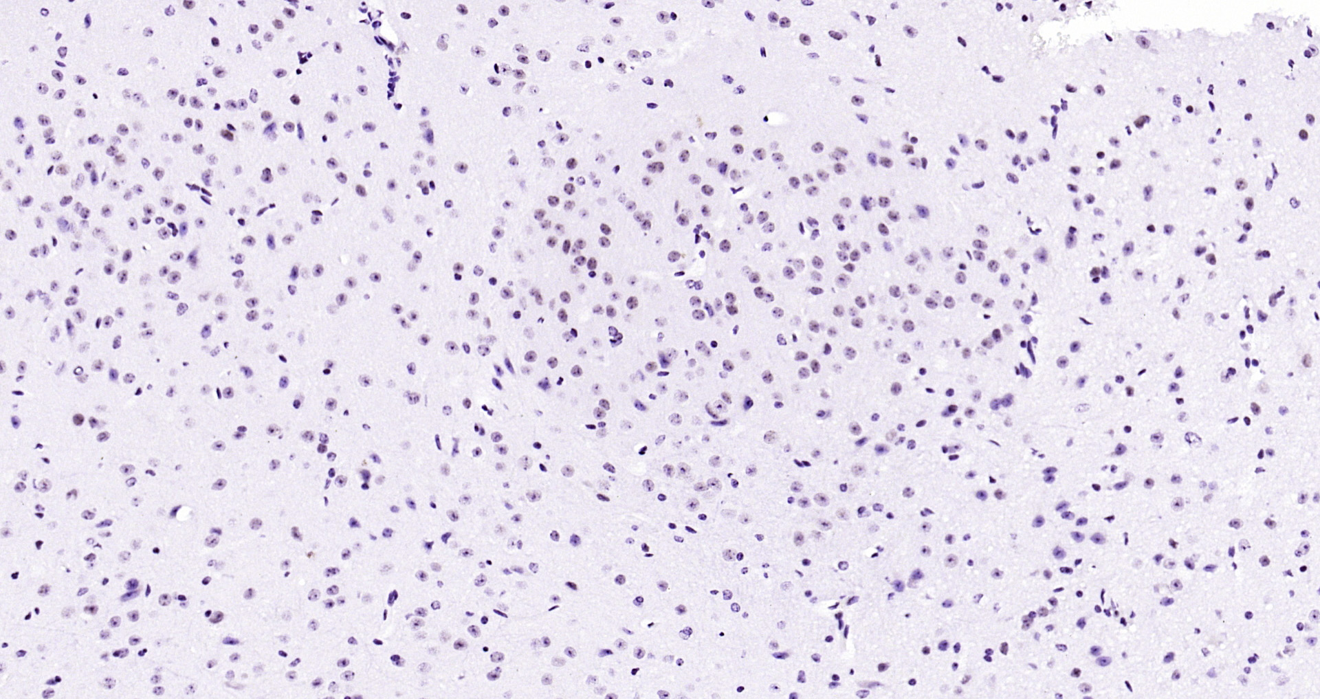
. API5 was detected in a paraffin-embedded section of Rat Brain tissue. Heat mediated antigen retrieval was performed in EDTA buffer (pH 8.0, epitope retrieval solution). The tissue section was blocked with 10% goat serum. The tissue section was then incubated with 1 microg/ml rabbit anti-API5 Antibody (PA1009) overnight at 4°C. Biotinylated goat anti-rabbit IgG was used as secondary antibody and incubated for 30 minutes at 37°C. The tissue section was developed using Strepavidin-Biotin-Complex (SABC) (Catalog # SA1022) with DAB as the chromogen.")
. API5 was detected in an immunocytochemical section of Hela cells. Enzyme antigen retrieval was performed using IHC enzyme antigen retrieval reagent (AR0022) for 15 mins. The cells were blocked with 10% goat serum. And then incubated with 1 microg/ml rabbit anti-API5 Antibody (PA1009) overnight at 4°C. Biotinylated goat anti-rabbit IgG was used as secondary antibody and incubated for 30 minutes at 37°C. The section was developed using Strepavidin-Biotin-Complex (SABC)(Catalog # SA1022) with DAB as the chromogen.")
. API5 was detected in a frozen section of Rat Cardiac Muscle tissue. The tissue section was blocked with 10% goat serum. The tissue section was then incubated with 1 microg/ml rabbit anti-API5 Antibody (PA1009) overnight at 4°C. Biotinylated goat anti-rabbit IgG was used as secondary antibody and incubated for 30 minutes at 37°C. The tissue section was developed using Strepavidin-Biotin-Complex (SABC) (Catalog # SA1022) with DAB as the chromogen.")
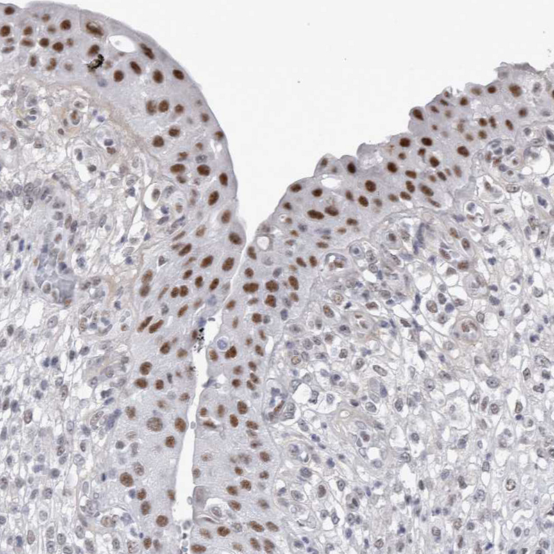
. API5 was detected in paraffin-embedded section of human placenta tissues. Heat mediated antigen retrieval was performed in citrate buffer (pH6, epitope retrieval solution) for 20 mins. The tissue section was blocked with 10% goat serum. The tissue section was then incubated with 1microg/ml rabbit anti-API5 Antibody (PA1009) overnight at 4°C. Biotinylated goat anti-rabbit IgG was used as secondary antibody and incubated for 30 minutes at 37°C. The tissue section was developed using Strepavidin-Biotin-Complex (SABC)(Catalog # SA1022) with DAB as the chromogen.")
. API5 was detected in paraffin-embedded section of rat intestines tissues. Heat mediated antigen retrieval was performed in citrate buffer (pH6, epitope retrieval solution) for 20 mins. The tissue section was blocked with 10% goat serum. The tissue section was then incubated with 1microg/ml rabbit anti-API5 Antibody (PA1009) overnight at 4°C. Biotinylated goat anti-rabbit IgG was used as secondary antibody and incubated for 30 minutes at 37°C. The tissue section was developed using Strepavidin-Biotin-Complex (SABC)(Catalog # SA1022) with DAB as the chromogen.")
. API5 was detected in paraffin-embedded section of mouse intestines tissues. Heat mediated antigen retrieval was performed in citrate buffer (pH6, epitope retrieval solution) for 20 mins. The tissue section was blocked with 10% goat serum. The tissue section was then incubated with 1microg/ml rabbit anti-API5 Antibody (PA1009) overnight at 4°C. Biotinylated goat anti-rabbit IgG was used as secondary antibody and incubated for 30 minutes at 37°C. The tissue section was developed using Strepavidin-Biotin-Complex (SABC)(Catalog # SA1022) with DAB as the chromogen.")
. API5 was detected in an immunocytochemical section of SK-OV-3 cells. Enzyme antigen retrieval was performed using IHC enzyme antigen retrieval reagent (AR0022) for 15 mins. The cells were blocked with 10% goat serum. And then incubated with 5 microg/mL rabbit anti-API5 Antibody (PA1009) overnight at 4°C. DyLight®488 Conjugated Goat Anti-Rabbit IgG (BA1127) was used as secondary antibody at 1:100 dilution and incubated for 30 minutes at 37°C. The section was counterstained with DAPI. Visualize using a fluorescence microscope and filter sets appropriate for the label used.")
Figure 1. Western blot analysis of API5 using anti-API5 antibody (PA1009). Electrophoresis was performed on a 5-20% SDS-PAGE gel at 70V (Stacking gel) / 90V (Resolving gel) for 2-3 hours. The sample well of each lane was loaded with 30 ug of sample under reducing conditions. Lane 1: Rat Cardiac Muscle Tissue Lysate Lane 2: Rat Brain Tissue Lysate, Lane 3: Rat Testis Tissue Lysate, Lane 4: Rat Placenta Tissue Lysate, Lane 5: MCF-7 Cell Lysate, Lane 6: HELA Cell Lysate, Lane 7: CEM Cell Lysate, Lane 8: SMMC Cell Lysate, Lane 9: COLO320 Cell Lysate. After electrophoresis, proteins were transferred to a nitrocellulose membrane at 150 mA for 50-90 minutes. Blocked the membrane with 5% non-fat milk/TBS for 1.5 hour at RT. The membrane was incubated with rabbit anti-API5 antigen affinity purified polyclonal antibody (Catalog # PA1009) at 0.5 microg/mL overnight at 4°C, then washed with TBS-0.1%Tween 3 times with 5 minutes each and probed with a goat anti-rabbit IgG-HRP secondary antibody at a dilution of 1:5000 for 1.5 hour at RT. The signal is developed using an Enhanced Chemiluminescent detection (ECL) kit (Catalog # EK1002) with Tanon 5200 system. A specific band was detected for API5 at approximately 59 kDa. The expected band size for API5 is at 59 kDa.
Anti-Apoptosis inhibitor 5/API5 Antibody Picoband(r)
PA1009-CARRIER-FREE
ApplicationsImmunoFluorescence, Western Blot, ImmunoCytoChemistry, ImmunoHistoChemistry, ImmunoHistoChemistry Frozen
Product group Antibodies
ReactivityHamster, Human, Mouse, Rat
TargetAPI5
Overview
- SupplierBoster Bio
- Product NameAnti-Apoptosis inhibitor 5/API5 Antibody Picoband(r)
- Delivery Days Customer9
- Application Supplier NoteTested Species: In-house tested species with positive results. Predicted Species: Species predicted to be fit for the product based on sequence similarities. By Heat: Boiling the paraffin sections in 10mM citrate buffer, pH6.0, for 20mins is required for the staining of formalin/paraffin sections. Other applications have not been tested. Optimal dilutions should be determined by end users.
- ApplicationsImmunoFluorescence, Western Blot, ImmunoCytoChemistry, ImmunoHistoChemistry, ImmunoHistoChemistry Frozen
- CertificationResearch Use Only
- ClonalityPolyclonal
- Concentration500 ug/ml
- Gene ID8539
- Target nameAPI5
- Target descriptionapoptosis inhibitor 5
- Target synonymsAAC-11, AAC11, apoptosis inhibitor 5, FIF, antiapoptosis clone 11 protein, cell migration-inducing gene 8 protein, fibroblast growth factor 2-interacting factor 2, migration-inducing protein MIG8
- HostRabbit
- IsotypeIgG
- Protein IDQ9BZZ5
- Protein NameApoptosis inhibitor 5
- Scientific DescriptionBoster Bio Anti-Apoptosis inhibitor 5/API5 Antibody catalog # PA1009. Tested in IF, IHC, IHC-F, ICC, WB applications. This antibody reacts with Human, Mouse, Rat. The brand Picoband indicates this is a premium antibody that guarantees superior quality, high affinity, and strong signals with minimal background in Western blot applications. Only our best-performing antibodies are designated as Picoband, ensuring unmatched performance.
- ReactivityHamster, Human, Mouse, Rat
- Storage Instruction-20°C,2°C to 8°C
- UNSPSC12352203
Related products
Product group Antibodies
API5 AntibodyCSB-PA000902
ApplicationsWestern Blot, ELISA, ImmunoHistoChemistry
ReactivityHuman, Mouse
TargetAPI5
- SizePrice
Product group Antibodies
API5 Polyclonal AntibodyCAC15024
ApplicationsImmunoFluorescence, Western Blot, ELISA
ReactivityMouse
TargetAPI5
- SizePrice
Product group Antibodies
Anti-API5 AntibodyA35992
ApplicationsWestern Blot, ELISA, ImmunoHistoChemistry
ReactivityHuman, Mouse
- SizePrice
Product group Antibodies
Anti-API5 AntibodyHPA026598
ApplicationsWestern Blot, ImmunoCytoChemistry, ImmunoHistoChemistry
ReactivityHuman, Mouse, Rat
TargetAPI5
- SizePrice
Product group Antibodies

API5 AntibodyLS-C406134
ApplicationsWestern Blot, ELISA
ReactivityHuman
TargetAPI5
- SizePrice
Product group Antibodies
API5 antibodyGTX38554
ApplicationsWestern Blot, ImmunoHistoChemistry, ImmunoHistoChemistry Paraffin
ReactivityHuman, Rat
TargetAPI5
- SizePrice
Product group Antibodies
Anti-API5 (C-term) Antibody102-23357
ApplicationsWestern Blot
TargetAPI5
- SizePrice
Product group Antibodies
API5 Polyclonal AntibodyBS-1252R
ApplicationsImmunoFluorescence, Western Blot, ImmunoHistoChemistry, ImmunoHistoChemistry Frozen, ImmunoHistoChemistry Paraffin
ReactivityBovine, Canine, Equine, Guinea Pig, Human, Mouse, Porcine, Rat
TargetAPI5
- SizePrice