


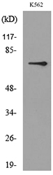
Figure 1. Western blot analysis of BMAL1/ARNTL using anti-BMAL1/ARNTL antibody (A00260-1). Electrophoresis was performed on a 5-20% SDS-PAGE gel at 70V (Stacking gel) / 90V (Resolving gel) for 2-3 hours. The sample well of each lane was loaded with 30 ug of sample under reducing conditions. Lane 1: human Hela whole cell lysates, Lane 2: human HT1080 whole cell lysates, Lane 3: human MCF-7 whole cell lysates, Lane 4: human SH-SY5Y whole cell lysates, Lane 5: rat brain tissue lysates, Lane 6: rat liver tissue lysates, Lane 7: mouse brain tissue lysates, Lane 8: mouse liver tissue lysates. After electrophoresis, proteins were transferred to a nitrocellulose membrane at 150 mA for 50-90 minutes. Blocked the membrane with 5% non-fat milk/TBS for 1.5 hour at RT. The membrane was incubated with rabbit anti-BMAL1/ARNTL antigen affinity purified polyclonal antibody (Catalog # A00260-1) at 0.5 microg/mL overnight at 4°C, then washed with TBS-0.1%Tween 3 times with 5 minutes each and probed with a goat anti-rabbit IgG-HRP secondary antibody at a dilution of 1:5000 for 1.5 hour at RT. The signal is developed using an Enhanced Chemiluminescent detection (ECL) kit (Catalog # EK1002) with Tanon 5200 system. A specific band was detected for BMAL1/ARNTL at approximately 75 kDa. The expected band size for BMAL1/ARNTL is at 69 kDa.
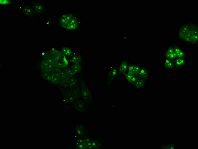
. Overlay histogram showing MCF-7 cells stained with A00260-1 (Blue line). To facilitate intracellular staining, cells were fixed with 4% paraformaldehyde and permeabilized with permeabilization buffer. The cells were blocked with 10% normal goat serum. And then incubated with rabbit anti-BMAL1/ARNTL Antibody (A00260-1, 1 microg/1x106 cells) for 30 min at 20°C. DyLight®488 conjugated goat anti-rabbit IgG (BA1127, 5-10 microg/1x106 cells) was used as secondary antibody for 30 minutes at 20°C. Isotype control antibody (Green line) was rabbit IgG (1 microg/1x106) used under the same conditions. Unlabelled sample without incubation with primary antibody and secondary antibody (Red line) was used as a blank control.")
Figure 1. Western blot analysis of BMAL1/ARNTL using anti-BMAL1/ARNTL antibody (A00260-1). Electrophoresis was performed on a 5-20% SDS-PAGE gel at 70V (Stacking gel) / 90V (Resolving gel) for 2-3 hours. The sample well of each lane was loaded with 30 ug of sample under reducing conditions. Lane 1: human Hela whole cell lysates, Lane 2: human HT1080 whole cell lysates, Lane 3: human MCF-7 whole cell lysates, Lane 4: human SH-SY5Y whole cell lysates, Lane 5: rat brain tissue lysates, Lane 6: rat liver tissue lysates, Lane 7: mouse brain tissue lysates, Lane 8: mouse liver tissue lysates. After electrophoresis, proteins were transferred to a nitrocellulose membrane at 150 mA for 50-90 minutes. Blocked the membrane with 5% non-fat milk/TBS for 1.5 hour at RT. The membrane was incubated with rabbit anti-BMAL1/ARNTL antigen affinity purified polyclonal antibody (Catalog # A00260-1) at 0.5 microg/mL overnight at 4°C, then washed with TBS-0.1%Tween 3 times with 5 minutes each and probed with a goat anti-rabbit IgG-HRP secondary antibody at a dilution of 1:5000 for 1.5 hour at RT. The signal is developed using an Enhanced Chemiluminescent detection (ECL) kit (Catalog # EK1002) with Tanon 5200 system. A specific band was detected for BMAL1/ARNTL at approximately 75 kDa. The expected band size for BMAL1/ARNTL is at 69 kDa.
Anti-BMAL1/ARNTL Antibody Picoband(r)
A00260-1-CARRIER-FREE
ApplicationsFlow Cytometry, Western Blot
Product group Antibodies
ReactivityHuman, Mouse, Rat
TargetBMAL1
Overview
- SupplierBoster Bio
- Product NameAnti-BMAL1/ARNTL Antibody Picoband(r)
- Delivery Days Customer9
- ApplicationsFlow Cytometry, Western Blot
- CertificationResearch Use Only
- ClonalityPolyclonal
- Concentration500 ug/ml
- Gene ID406
- Target nameBMAL1
- Target descriptionbasic helix-loop-helix ARNT like 1
- Target synonymsARNTL, ARNTL1, BMAL1c, JAP3, MOP3, PASD3, TIC, bHLHe5, basic helix-loop-helix ARNT-like protein 1, ARNT-like protein 1, brain and muscle, PAS domain containing 3, PAS domain-containing protein 3, aryl hydrocarbon receptor nuclear translocator like, aryl hydrocarbon receptor nuclear translocator-like protein 1, bHLH-PAS protein JAP3, basic helix-loop-helix family member e5, basic-helix-loop-helix-PAS orphan MOP3, basic-helix-loop-helix-PAS protein MOP3, brain and muscle ARNT-like 1, class E basic helix-loop-helix protein 5, member of PAS protein 3, member of PAS superfamily 3, mutant basic helix-loop-helix ARNT-like protein 1, testis tissue sperm-binding protein Li 50e
- HostRabbit
- IsotypeIgG
- Protein IDO00327
- Protein NameBasic helix-loop-helix ARNT-like protein 1
- Scientific DescriptionBoster Bio Anti-BMAL1/ARNTL Antibody Picoband® catalog # A00260-1. Tested in Flow Cytometry, WB applications. This antibody reacts with Human, Mouse, Rat. The brand Picoband indicates this is a premium antibody that guarantees superior quality, high affinity, and strong signals with minimal background in Western blot applications. Only our best-performing antibodies are designated as Picoband, ensuring unmatched performance.
- ReactivityHuman, Mouse, Rat
- Storage Instruction-20°C,2°C to 8°C
- UNSPSC12352203
Related products
Product group Antibodies
Anti-BMAL1 AntibodyA97688
ApplicationsWestern Blot, ELISA
ReactivityHuman, Mouse, Rat
- SizePrice
Product group Antibodies
BMAL1 Polyclonal Antibodybs-3750R
ApplicationsFlow Cytometry, ImmunoFluorescence, Western Blot, ELISA, ImmunoCytoChemistry, ImmunoHistoChemistry, ImmunoHistoChemistry Frozen, ImmunoHistoChemistry Paraffin
ReactivityBovine, Canine, Equine, Human, Mouse, Porcine, Rat, Sheep
TargetBMAL1
- SizePrice
Product group Antibodies
ARNTL AntibodyCSB-PA002123LA01HU
ApplicationsImmunoFluorescence, ELISA
ReactivityHuman
TargetBMAL1
- SizePrice
Product group Antibodies

Goat anti-BMAL1 / ARNTLEB09178
ApplicationsWestern Blot, ELISA
ReactivityBovine, Canine, Human, Mouse, Porcine, Rat
TargetBMAL1
- SizePrice
Product group Antibodies

ARNTL / BMAL1 AntibodyLS-C401453
ApplicationsWestern Blot, ELISA
ReactivityHuman, Mouse, Rat
TargetBMAL1
- SizePrice

Product group Antibodies
Anti-ARNTL AntibodyHPA050938
ApplicationsImmunoCytoChemistry
ReactivityHuman
TargetBMAL1
- SizePrice
![Treated 293T whole cell extracts (30 μg) were separated by 7.5% SDS-PAGE, and the membranes were blotted with BMAL1 antibody [N1N3] (GTX105060) and PER2 antibody (GTX129688) diluted at 1:500. The HRP-conjugated anti-rabbit IgG antibody (GTX213110-01) was used to detect the primary antibody.](https://www.genetex.com/upload/website/prouct_img/normal/GTX105060/GTX105060_39876_20191025_WB_treatment_Serumshock_watermark_w_23060120_168.webp)
Product group Antibodies
BMAL1 antibody [N1N3]GTX105060
ApplicationsImmunoFluorescence, Western Blot, ImmunoCytoChemistry
ReactivityHuman, Mouse
TargetBMAL1
- SizePrice
Product group Antibodies
ApplicationsWestern Blot, ELISA
ReactivityRat
TargetBMAL1
- SizePrice