


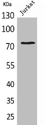
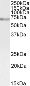
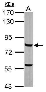
Figure 1. Western blot analysis of c-Myb using anti-c-Myb antibody (PB9288). Electrophoresis was performed on a 5-20% SDS-PAGE gel at 70V (Stacking gel) / 90V (Resolving gel) for 2-3 hours. The sample well of each lane was loaded with 30 ug of sample under reducing conditions. Lane 1: human K562 whole cell lysates, Lane 2: human MCF-7 whole cell lysates, Lane 3: human HEK293 whole cell lysates. After electrophoresis, proteins were transferred to a nitrocellulose membrane at 150 mA for 50-90 minutes. Blocked the membrane with 5% non-fat milk/TBS for 1.5 hour at RT. The membrane was incubated with rabbit anti-c-Myb antigen affinity purified polyclonal antibody (Catalog # PB9288) at 0.5 microg/mL overnight at 4°C, then washed with TBS-0.1%Tween 3 times with 5 minutes each and probed with a goat anti-rabbit IgG-HRP secondary antibody at a dilution of 1:5000 for 1.5 hour at RT. The signal is developed using an Enhanced Chemiluminescent detection (ECL) kit (Catalog # EK1002) with Tanon 5200 system. A specific band was detected for c-Myb at approximately 74 kDa. The expected band size for c-Myb is at 74 kDa.
Figure 1. Western blot analysis of c-Myb using anti-c-Myb antibody (PB9288). Electrophoresis was performed on a 5-20% SDS-PAGE gel at 70V (Stacking gel) / 90V (Resolving gel) for 2-3 hours. The sample well of each lane was loaded with 30 ug of sample under reducing conditions. Lane 1: human K562 whole cell lysates, Lane 2: human MCF-7 whole cell lysates, Lane 3: human HEK293 whole cell lysates. After electrophoresis, proteins were transferred to a nitrocellulose membrane at 150 mA for 50-90 minutes. Blocked the membrane with 5% non-fat milk/TBS for 1.5 hour at RT. The membrane was incubated with rabbit anti-c-Myb antigen affinity purified polyclonal antibody (Catalog # PB9288) at 0.5 microg/mL overnight at 4°C, then washed with TBS-0.1%Tween 3 times with 5 minutes each and probed with a goat anti-rabbit IgG-HRP secondary antibody at a dilution of 1:5000 for 1.5 hour at RT. The signal is developed using an Enhanced Chemiluminescent detection (ECL) kit (Catalog # EK1002) with Tanon 5200 system. A specific band was detected for c-Myb at approximately 74 kDa. The expected band size for c-Myb is at 74 kDa.
Anti-c-Myb Antibody Picoband(r)
PB9288-CARRIER-FREE
ApplicationsFlow Cytometry, ImmunoFluorescence, Western Blot, ImmunoHistoChemistry
Product group Antibodies
ReactivityBovine, Human, Monkey, Rabbit
TargetMYB
Overview
- SupplierBoster Bio
- Product NameAnti-c-Myb Antibody Picoband(r)
- Delivery Days Customer9
- Application Supplier NoteWB: The detection limit for c-Myb is approximately 0.25ng/lane under reducing conditions. Tested Species: In-house tested species with positive results. Other applications have not been tested. Optimal dilutions should be determined by end users.
- ApplicationsFlow Cytometry, ImmunoFluorescence, Western Blot, ImmunoHistoChemistry
- CertificationResearch Use Only
- ClonalityPolyclonal
- Concentration500 ug/ml
- Gene ID4602
- Target nameMYB
- Target descriptionMYB proto-oncogene, transcription factor
- Target synonymsCmyb, c-myb, c-myb_CDS, efg, transcriptional activator Myb, MYB-GATA1 fusion protein, oncogene AMV, proto-oncogene c-Myb, v-myb avian myeloblastosis viral oncogene homolog
- HostRabbit
- IsotypeIgG
- Protein IDP10242
- Protein NameTranscriptional activator Myb
- Scientific DescriptionBoster Bio Anti-c-Myb Antibody Picoband® catalog # PB9288. Tested in Flow Cytometry, IHC, IF, WB applications. This antibody reacts with Human. The brand Picoband indicates this is a premium antibody that guarantees superior quality, high affinity, and strong signals with minimal background in Western blot applications. Only our best-performing antibodies are designated as Picoband, ensuring unmatched performance.
- ReactivityBovine, Human, Monkey, Rabbit
- Storage Instruction-20°C,2°C to 8°C
- UNSPSC12352203
Related products
Product group Antibodies
MYB AntibodyCSB-PA005513
ApplicationsWestern Blot, ELISA
ReactivityHuman, Mouse
TargetMYB
- SizePrice
Product group Antibodies
Anti-c-Myb AntibodyA82604
ApplicationsWestern Blot, ELISA, ImmunoHistoChemistry
ReactivityHuman
- SizePrice
Product group Antibodies

Myb Sumoylation Site AntibodyABX031602
ApplicationsWestern Blot, ELISA, ImmunoHistoChemistry
- SizePrice
Product group Antibodies

MYB / c-Myb AntibodyLS-C748801
ApplicationsWestern Blot
ReactivityHuman
TargetMYB
- SizePrice
Product group Antibodies

Goat anti-c-Myb (aa281-294)EB07870
ApplicationsWestern Blot, ELISA, ImmunoHistoChemistry
ReactivityCanine, Human, Mouse, Rat
TargetMYB
- SizePrice

Product group Antibodies
Anti-MYB AntibodyHPA071605
ApplicationsChIP Chromatin ImmunoPrecipitation, ImmunoCytoChemistry
ReactivityHuman
TargetMYB
- SizePrice
Product group Antibodies
References
MYBL2 Polyclonal Antibodybs-5960R
ApplicationsImmunoFluorescence, Western Blot, ELISA, ImmunoCytoChemistry, ImmunoHistoChemistry, ImmunoHistoChemistry Frozen, ImmunoHistoChemistry Paraffin
ReactivityBovine, Canine, Equine, Human, Mouse, Porcine, Rabbit, Rat
TargetMYB
- SizePrice
Product group Antibodies
ApplicationsImmunoPrecipitation, Western Blot, ImmunoCytoChemistry, ImmunoHistoChemistry
ReactivityMouse
TargetMYB
- SizePrice
Product group Antibodies
c-Myb antibody [N2C1], InternalGTX102305
ApplicationsWestern Blot
ReactivityHuman
TargetMYB
- SizePrice