

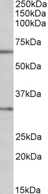


Anti-Caspase-3 Antibody
A83337
ApplicationsImmunoFluorescence, Western Blot, ELISA, ImmunoCytoChemistry
Product group Antibodies
ReactivityHuman, Mouse, Rat
Overview
- SupplierAntibodies.com
- Product NameAnti-Caspase-3 Antibody
- Delivery Days Customer7
- ApplicationsImmunoFluorescence, Western Blot, ELISA, ImmunoCytoChemistry
- CertificationResearch Use Only
- ClonalityPolyclonal
- Concentration500 ug/ml
- ConjugateUnconjugated
- HostGoat
- IsotypeIgG
- Scientific DescriptionGoat polyclonal antibody to Caspase-3.
- ReactivityHuman, Mouse, Rat
- UNSPSC12352203
Related products
Product group Antibodies
Anti-CASP3 (138-157aa) Antibody130-10514
ApplicationsELISA
ReactivityHuman
TargetCASP3
- SizePrice
Product group Antibodies

Anti-Caspase-3 [4F-6]AB04219-1.1
ApplicationsWestern Blot, ELISA, Neutralisation/Blocking
ReactivityHuman
TargetCASP3
- SizePrice

Product group Antibodies
Anti-Caspase-3(p17)/CASP3 Antibody Picoband(r)A00334-2-CARRIER-FREE
ApplicationsFlow Cytometry, ImmunoFluorescence, Western Blot, ImmunoCytoChemistry
ReactivityHuman
TargetCASP3
- SizePrice

Product group Antibodies
References
Caspase-3 Polyclonal AntibodyBS-0081R
ApplicationsWestern Blot, ELISA, ImmunoHistoChemistry, ImmunoHistoChemistry Paraffin
ReactivityBovine, Chicken, Human, Monkey, Mouse, Porcine, Rabbit, Rat, Sheep, Other Species
TargetCASP3
- SizePrice
Product group Antibodies
CASP3 Monoclonal AntibodyCSB-MA080226
ApplicationsWestern Blot, ELISA, ImmunoHistoChemistry
ReactivityHuman, Mouse, Rat
TargetCASP3
- SizePrice
Product group Antibodies
References

Goat anti-Caspase 3EB07286
ApplicationsImmunoFluorescence, Western Blot, ELISA, ImmunoCytoChemistry
ReactivityCanine, Human, Mouse, Rat
TargetCASP3
- SizePrice
Product group Antibodies
ApplicationsWestern Blot, ImmunoHistoChemistry
ReactivityPorcine, Rat
TargetCASP3
- SizePrice
Product group Antibodies

ApplicationsELISA
ReactivityHuman
TargetCASP3
- SizePrice

Product group Antibodies
Anti-CASP3 AntibodyHPA002643
ApplicationsWestern Blot, ImmunoCytoChemistry, ImmunoHistoChemistry
ReactivityHuman
TargetCASP3
- SizePrice