

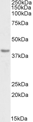
Anti-PD-L1 Antibody (Biotin)
A82539
ApplicationsWestern Blot, ELISA
Product group Antibodies
ReactivityHuman
Overview
- SupplierAntibodies.com
- Product NameAnti-PD-L1 Antibody (Biotin)
- Delivery Days Customer7
- ApplicationsWestern Blot, ELISA
- CertificationResearch Use Only
- ClonalityPolyclonal
- Concentration500 ug/ml
- ConjugateBiotin
- HostGoat
- IsotypeIgG
- Scientific DescriptionGoat polyclonal antibody to PD-L1 (Biotin).
- ReactivityHuman
- UNSPSC12352203
Related products
Product group Antibodies
anti-PD-L1 (human), mAb (AG-IHC411)AG-20B-6022
ApplicationsELISA, ImmunoHistoChemistry
ReactivityHuman
TargetCD274
- SizePrice
Product group Antibodies

Anti-PD-L1 [mAb #18 (PD-L1.A)]Ab03505-1.1
ApplicationsWestern Blot, Neutralisation/Blocking, Other Application
ReactivityHuman
TargetCD274
- SizePrice
Product group Antibodies
Anti-PD-L1 Antibody130-10633
ApplicationsELISA
ReactivityHuman
TargetCD274
- SizePrice
Product group Antibodies

ApplicationsFlow Cytometry, ImmunoFluorescence, ImmunoHistoChemistry
ReactivityMouse
TargetCD274
- SizePrice
Product group Antibodies
Anti-PD-L1/B7-H1/CD274 Antibody Picoband(r)A00109-CARRIER-FREE
ApplicationsWestern Blot
ReactivityHuman
TargetCD274
- SizePrice
Product group Antibodies
PD-L1 Polyclonal AntibodyBS-22022R
ApplicationsImmunoFluorescence, Western Blot, ELISA, ImmunoCytoChemistry, ImmunoHistoChemistry, ImmunoHistoChemistry Frozen, ImmunoHistoChemistry Paraffin
ReactivityHuman, Mouse, Rat
TargetCD274
- SizePrice
Product group Antibodies

ApplicationsWestern Blot, ELISA
ReactivityHuman
TargetCD274
- SizePrice
Product group Antibodies
CD274 AntibodyCSB-PA878942ESR1HU
ApplicationsELISA, ImmunoHistoChemistry
ReactivityHuman
TargetCD274
- SizePrice
Product group Antibodies

CD274, PD-L1CD274-0.5
ApplicationsImmunoHistoChemistry, ImmunoHistoChemistry Frozen, ImmunoHistoChemistry Paraffin
ReactivityHuman
TargetCD274
- SizePrice
Product group Antibodies
ApplicationsFlow Cytometry
TargetCD274
- SizePrice