

Figure 2. IHC analysis of CHAT using anti-CHAT antibody (A01192-3). CHAT was detected in paraffin-embedded section of mouse brain tissues. Heat mediated antigen retrieval was performed in citrate buffer (pH6, epitope retrieval solution) for 20 mins. The tissue section was blocked with 10% goat serum. The tissue section was then incubated with 1microg/ml rabbit anti-CHAT Antibody (A01192-3) overnight at 4°C. Biotinylated goat anti-rabbit IgG was used as secondary antibody and incubated for 30 minutes at 37°C. The tissue section was developed using Strepavidin-Biotin-Complex (SABC)(Catalog # SA1022) with DAB as the chromogen.
. CHAT was detected in paraffin-embedded section of mouse brain tissues. Heat mediated antigen retrieval was performed in citrate buffer (pH6, epitope retrieval solution) for 20 mins. The tissue section was blocked with 10% goat serum. The tissue section was then incubated with 1microg/ml rabbit anti-CHAT Antibody (A01192-3) overnight at 4°C. Biotinylated goat anti-rabbit IgG was used as secondary antibody and incubated for 30 minutes at 37°C. The tissue section was developed using Strepavidin-Biotin-Complex (SABC)(Catalog # SA1022) with DAB as the chromogen.")
. CHAT was detected in paraffin-embedded section of rat brain tissues. Heat mediated antigen retrieval was performed in citrate buffer (pH6, epitope retrieval solution) for 20 mins. The tissue section was blocked with 10% goat serum. The tissue section was then incubated with 1microg/ml rabbit anti-CHAT Antibody (A01192-3) overnight at 4°C. Biotinylated goat anti-rabbit IgG was used as secondary antibody and incubated for 30 minutes at 37°C. The tissue section was developed using Strepavidin-Biotin-Complex (SABC)(Catalog # SA1022) with DAB as the chromogen.")
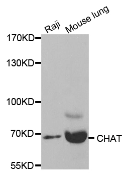
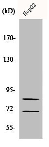
. Electrophoresis was performed on a 5-20% SDS-PAGE gel at 70V (Stacking gel) / 90V (Resolving gel) for 2-3 hours. The sample well of each lane was loaded with 50ug of sample under reducing conditions. Lane 1: rat testis tissue lysates, Lane 2: mouse testis tissue lysates. After Electrophoresis, proteins were transferred to a Nitrocellulose membrane at 150mA for 50-90 minutes. Blocked the membrane with 5% Non-fat Milk/ TBS for 1.5 hour at RT. The membrane was incubated with rabbit anti-CHAT antigen affinity purified polyclonal antibody (Catalog # A01192-3) at 0.5 microg/mL overnight at 4°C, then washed with TBS-0.1%Tween 3 times with 5 minutes each and probed with a goat anti-rabbit IgG-HRP secondary antibody at a dilution of 1:10000 for 1.5 hour at RT. The signal is developed using an Enhanced Chemiluminescent detection (ECL) kit (Catalog # EK1002) with Tanon 5200 system. A specific band was detected for CHAT at approximately 83KD. The expected band size for CHAT is at 83KD.")
Figure 2. IHC analysis of CHAT using anti-CHAT antibody (A01192-3). CHAT was detected in paraffin-embedded section of mouse brain tissues. Heat mediated antigen retrieval was performed in citrate buffer (pH6, epitope retrieval solution) for 20 mins. The tissue section was blocked with 10% goat serum. The tissue section was then incubated with 1microg/ml rabbit anti-CHAT Antibody (A01192-3) overnight at 4°C. Biotinylated goat anti-rabbit IgG was used as secondary antibody and incubated for 30 minutes at 37°C. The tissue section was developed using Strepavidin-Biotin-Complex (SABC)(Catalog # SA1022) with DAB as the chromogen.
Anti-Choline Acetyltransferase/CHAT Antibody Picoband(r)
A01192-3-CARRIER-FREE
ApplicationsWestern Blot, ImmunoHistoChemistry
Product group Antibodies
ReactivityHuman, Mouse, Rat
TargetCHAT
Overview
- SupplierBoster Bio
- Product NameAnti-Choline Acetyltransferase/CHAT Antibody Picoband(r)
- Delivery Days Customer9
- ApplicationsWestern Blot, ImmunoHistoChemistry
- CertificationResearch Use Only
- ClonalityPolyclonal
- Concentration500 ug/ml
- Gene ID1103
- Target nameCHAT
- Target descriptioncholine O-acetyltransferase
- Target synonymsCHOACTASE, CMS1A, CMS1A2, CMS6, choline O-acetyltransferase, acetyl CoA:choline O-acetyltransferase, choline acetylase
- HostRabbit
- IsotypeIgG
- Protein IDP28329
- Protein NameCholine O-acetyltransferase
- Scientific DescriptionBoster Bio Anti-Choline Acetyltransferase/CHAT Antibody Picoband® catalog # A01192-3. Tested in IHC, WB applications. This antibody reacts with Human, Mouse, Rat. The brand Picoband indicates this is a premium antibody that guarantees superior quality, high affinity, and strong signals with minimal background in Western blot applications. Only our best-performing antibodies are designated as Picoband, ensuring unmatched performance.
- ReactivityHuman, Mouse, Rat
- Storage Instruction-20°C,2°C to 8°C
- UNSPSC12352203
Related products
Product group Antibodies
Anti-CHAT AntibodyA35519
ApplicationsImmunoFluorescence, Western Blot, ImmunoHistoChemistry
ReactivityHuman, Mouse, Rat
- SizePrice
Product group Antibodies
Anti-CHAT Antibody144-02495
ApplicationsWestern Blot, ImmunoHistoChemistry
ReactivityHuman, Mouse, Rat
TargetCHAT
- SizePrice

Product group Antibodies
Anti-CHAT AntibodyAMAB91129
ApplicationsImmunoHistoChemistry
ReactivityHuman, Mouse, Rat
TargetCHAT
- SizePrice
Product group Antibodies
References
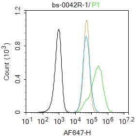
ChAT Polyclonal AntibodyBS-0042R
ApplicationsFlow Cytometry, ImmunoFluorescence, ELISA, ImmunoCytoChemistry, ImmunoHistoChemistry, ImmunoHistoChemistry Frozen, ImmunoHistoChemistry Paraffin
ReactivityHuman
TargetCHAT
- SizePrice
Product group Antibodies
CHAT AntibodyCSB-PA001637
ApplicationsWestern Blot, ELISA
ReactivityHuman, Mouse, Rat
TargetCHAT
- SizePrice
Product group Antibodies

ApplicationsWestern Blot, ELISA
ReactivityBovine, Canine, Human, Porcine
TargetCHAT
- SizePrice
Product group Antibodies
Chat Polyclonal AntibodyCAC07701
ApplicationsImmunoFluorescence, Western Blot, ELISA, ImmunoHistoChemistry
ReactivityMouse
TargetCHAT
- SizePrice
![Choline Acetyltransferase antibody [N1N3] detects Choline Acetyltransferase protein at cytoplasm and nucleus by immunohistochemical analysis. Sample: Paraffin-embedded mouse eye. Green: Choline Acetyltransferase stained by Choline Acetyltransferase antibody [N1N3] (GTX113164) diluted at 1:615. Red: beta Tubulin 3/ Tuj1, a Cytoskeleton marker, stained by beta Tubulin 3/ Tuj1 antibody [GT11710] (GTX631836) diluted at 1:500. Blue: Fluoroshield with DAPI (GTX30920). Antigen Retrieval: Citrate buffer, pH 6.0, 15 min](https://www.genetex.com/upload/website/prouct_img/normal/GTX113164/GTX113164_44720_20220715_IHC-P_M_1_22071823_849.webp)
Product group Antibodies
ApplicationsImmunoFluorescence, Western Blot, ImmunoCytoChemistry, ImmunoHistoChemistry, ImmunoHistoChemistry Frozen, ImmunoHistoChemistry Paraffin
ReactivityHuman, Mouse, Rat
TargetCHAT
- SizePrice