

Choline Acetyltransferase antibody [N1N3] detects Choline Acetyltransferase protein at cytoplasm and nucleus by immunohistochemical analysis. Sample: Paraffin-embedded mouse eye. Green: Choline Acetyltransferase stained by Choline Acetyltransferase antibody [N1N3] (GTX113164) diluted at 1:615. Red: beta Tubulin 3/ Tuj1, a Cytoskeleton marker, stained by beta Tubulin 3/ Tuj1 antibody [GT11710] (GTX631836) diluted at 1:500. Blue: Fluoroshield with DAPI (GTX30920). Antigen Retrieval: Citrate buffer, pH 6.0, 15 min
![Non-transfected (–) and transfected (+) 293T whole cell extracts (30 μg) were separated by 7.5% SDS-PAGE, and the membrane was blotted with Choline Acetyltransferase antibody [N1N3] (GTX113164) diluted at 1:5000. The HRP-conjugated anti-rabbit IgG antibody (GTX213110-01) was used to detect the primary antibody.](https://www.genetex.com/upload/website/prouct_img/normal/GTX113164/GTX113164_43173_20180413_WB_B_w_23060500_521.webp "Non-transfected (–) and transfected (+) 293T whole cell extracts (30 μg) were separated by 7.5% SDS-PAGE, and the membrane was blotted with Choline Acetyltransferase antibody [N1N3] (GTX113164) diluted at 1:5000. The HRP-conjugated anti-rabbit IgG antibody (GTX213110-01) was used to detect the primary antibody.")
![Choline Acetyltransferase antibody [N1N3] detects Choline Acetyltransferase protein by immunohistochemical analysis. Sample: Frozen-sectioned mouse spinal cord. Red: Choline Acetyltransferase stained by Choline Acetyltransferase antibody [N1N3] (GTX113164) diluted at 1:300. Blue: Hoechst 33342 staining.](https://www.genetex.com/upload/website/prouct_img/normal/GTX113164/GTX113164_43629_20200303_IHC-Fr_M_w_23060501_332.webp "Choline Acetyltransferase antibody [N1N3] detects Choline Acetyltransferase protein by immunohistochemical analysis. Sample: Frozen-sectioned mouse spinal cord. Red: Choline Acetyltransferase stained by Choline Acetyltransferase antibody [N1N3] (GTX113164) diluted at 1:300. Blue: Hoechst 33342 staining.")
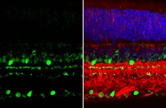
![Choline Acetyltransferase antibody [N1N3] detects Choline Acetyltransferase protein in the amacrine cells by immunohistochemical analysis. Sample: Frozen sectioned adult mouse retina. Green: Choline Acetyltransferase protein stained by Choline Acetyltransferase antibody [N1N3] (GTX113164) diluted at 1:250. Red: Protein kinase C alpha staining. Blue: Fluoroshield with DAPI (GTX30920).
Antigen Retrieval: Citrate buffer, pH 6.0, 15 min](https://www.genetex.com/upload/website/prouct_img/normal/GTX113164/GTX113164_40086_20160808_IHC-Fr_w_23060501_225.webp "Choline Acetyltransferase antibody [N1N3] detects Choline Acetyltransferase protein in the amacrine cells by immunohistochemical analysis. Sample: Frozen sectioned adult mouse retina. Green: Choline Acetyltransferase protein stained by Choline Acetyltransferase antibody [N1N3] (GTX113164) diluted at 1:250. Red: Protein kinase C alpha staining. Blue: Fluoroshield with DAPI (GTX30920).
Antigen Retrieval: Citrate buffer, pH 6.0, 15 min")
![Choline Acetyltransferase antibody [N1N3] detects Choline Acetyltransferase protein by immunofluorescent analysis. Sample: DIV14 rat E18 primary cortical neurons were fixed in 4% paraformaldehyde at RT for 15 min. Green: Choline Acetyltransferase protein stained by Choline Acetyltransferase antibody [N1N3] (GTX113164) diluted at 1:500. Red: beta Tubulin 3/ Tuj1, stained by beta Tubulin 3/ Tuj1 antibody [GT1338] (GTX631831) diluted at 1:500. Blue: Fluoroshield with DAPI (GTX30920).](https://www.genetex.com/upload/website/prouct_img/normal/GTX113164/GTX113164_40086_20170719_IFA_R_w_23060501_177.webp "Choline Acetyltransferase antibody [N1N3] detects Choline Acetyltransferase protein by immunofluorescent analysis. Sample: DIV14 rat E18 primary cortical neurons were fixed in 4% paraformaldehyde at RT for 15 min. Green: Choline Acetyltransferase protein stained by Choline Acetyltransferase antibody [N1N3] (GTX113164) diluted at 1:500. Red: beta Tubulin 3/ Tuj1, stained by beta Tubulin 3/ Tuj1 antibody [GT1338] (GTX631831) diluted at 1:500. Blue: Fluoroshield with DAPI (GTX30920).")
![Mouse tissue extract (50 μg) was separated by 7.5% SDS-PAGE, and the membrane was blotted with Choline Acetyltransferase antibody [N1N3] (GTX113164) diluted at 1:1000. The HRP-conjugated anti-rabbit IgG antibody (GTX213110-01) was used to detect the primary antibody.](https://www.genetex.com/upload/website/prouct_img/normal/GTX113164/GTX113164_44685_20220520_WB_M_spinalcord_24012217_627.webp "Mouse tissue extract (50 μg) was separated by 7.5% SDS-PAGE, and the membrane was blotted with Choline Acetyltransferase antibody [N1N3] (GTX113164) diluted at 1:1000. The HRP-conjugated anti-rabbit IgG antibody (GTX213110-01) was used to detect the primary antibody.")
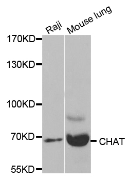
![Various tissue extracts (50 μg) were separated by 7.5% SDS-PAGE, and the membrane was blotted with Choline Acetyltransferase antibody [N1N3] (GTX113164) diluted at 1:500. The HRP-conjugated anti-rabbit IgG antibody (GTX213110-01) was used to detect the primary antibody.](https://www.genetex.com/upload/website/prouct_img/normal/GTX113164/GTX113164_44720_20220624_WB_M_R_24070822_520.webp "Various tissue extracts (50 μg) were separated by 7.5% SDS-PAGE, and the membrane was blotted with Choline Acetyltransferase antibody [N1N3] (GTX113164) diluted at 1:500. The HRP-conjugated anti-rabbit IgG antibody (GTX213110-01) was used to detect the primary antibody.")
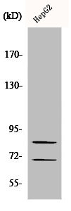
![Whole cell extract (30 μg) was separated by 7.5% SDS-PAGE, and the membrane was blotted with Choline Acetyltransferase antibody [N1N3] (GTX113164) diluted at 1:500. The HRP-conjugated anti-rabbit IgG antibody (GTX213110-01) was used to detect the primary antibody.](https://www.genetex.com/upload/website/prouct_img/normal/GTX113164/GTX113164_44720_20220624_WB_24070822_775.webp "Whole cell extract (30 μg) was separated by 7.5% SDS-PAGE, and the membrane was blotted with Choline Acetyltransferase antibody [N1N3] (GTX113164) diluted at 1:500. The HRP-conjugated anti-rabbit IgG antibody (GTX213110-01) was used to detect the primary antibody.")
Choline Acetyltransferase antibody [N1N3] detects Choline Acetyltransferase protein at cytoplasm and nucleus by immunohistochemical analysis. Sample: Paraffin-embedded mouse eye. Green: Choline Acetyltransferase stained by Choline Acetyltransferase antibody [N1N3] (GTX113164) diluted at 1:615. Red: beta Tubulin 3/ Tuj1, a Cytoskeleton marker, stained by beta Tubulin 3/ Tuj1 antibody [GT11710] (GTX631836) diluted at 1:500. Blue: Fluoroshield with DAPI (GTX30920). Antigen Retrieval: Citrate buffer, pH 6.0, 15 min
Choline Acetyltransferase antibody [N1N3]
GTX113164
ApplicationsImmunoFluorescence, Western Blot, ImmunoCytoChemistry, ImmunoHistoChemistry, ImmunoHistoChemistry Frozen, ImmunoHistoChemistry Paraffin
Product group Antibodies
ReactivityHuman, Mouse, Rat
TargetCHAT
Overview
- SupplierGeneTex
- Product NameCholine Acetyltransferase antibody [N1N3]
- Delivery Days Customer9
- Application Supplier NoteWB: 1:500-1:3000. ICC/IF: 1:100-1:1000. IHC-P: 1:100-1:1000. IHC-Fr: 1:100-1:1000. *Optimal dilutions/concentrations should be determined by the researcher.Not tested in other applications.
- ApplicationsImmunoFluorescence, Western Blot, ImmunoCytoChemistry, ImmunoHistoChemistry, ImmunoHistoChemistry Frozen, ImmunoHistoChemistry Paraffin
- CertificationResearch Use Only
- ClonalityPolyclonal
- Concentration0.15 mg/ml
- ConjugateUnconjugated
- Gene ID1103
- Target nameCHAT
- Target descriptioncholine O-acetyltransferase
- Target synonymsCHOACTASE, CMS1A, CMS1A2, CMS6, choline O-acetyltransferase, acetyl CoA:choline O-acetyltransferase, choline acetylase
- HostRabbit
- IsotypeIgG
- Protein IDP28329
- Protein NameCholine O-acetyltransferase
- Scientific DescriptionThis gene encodes an enzyme which catalyzes the biosynthesis of the neurotransmitter acetylcholine. This gene product is a characteristic feature of cholinergic neurons, and changes in these neurons may explain some of the symptoms of Alzheimer disease. Mutations in this gene are associated with congenital myasthenic syndrome associated with episodic apnea. Multiple transcript variants encoding different isoforms have been found for this gene, and some of these variants have been shown to encode more than one isoform. [provided by RefSeq]
- ReactivityHuman, Mouse, Rat
- Storage Instruction-20°C or -80°C,2°C to 8°C
- UNSPSC41116161
Datasheet
Related products
Product group Antibodies
Anti-CHAT AntibodyA35519
ApplicationsImmunoFluorescence, Western Blot, ImmunoHistoChemistry
ReactivityHuman, Mouse, Rat
- SizePrice

Product group Antibodies
Anti-Choline Acetyltransferase/CHAT Antibody Picoband(r)A01192-3-CARRIER-FREE
ApplicationsWestern Blot, ImmunoHistoChemistry
ReactivityHuman, Mouse, Rat
TargetCHAT
- SizePrice
Product group Antibodies
Anti-CHAT Antibody144-02495
ApplicationsWestern Blot, ImmunoHistoChemistry
ReactivityHuman, Mouse, Rat
TargetCHAT
- SizePrice

Product group Antibodies
Anti-CHAT AntibodyAMAB91129
ApplicationsImmunoHistoChemistry
ReactivityHuman, Mouse, Rat
TargetCHAT
- SizePrice
Product group Antibodies
References
ChAT Polyclonal AntibodyBS-0042R
ApplicationsFlow Cytometry, ImmunoFluorescence, ELISA, ImmunoCytoChemistry, ImmunoHistoChemistry, ImmunoHistoChemistry Frozen, ImmunoHistoChemistry Paraffin
ReactivityHuman
TargetCHAT
- SizePrice
Product group Antibodies
CHAT AntibodyCSB-PA001637
ApplicationsWestern Blot, ELISA
ReactivityHuman, Mouse, Rat
TargetCHAT
- SizePrice
Product group Antibodies

ApplicationsWestern Blot, ELISA
ReactivityBovine, Canine, Human, Porcine
TargetCHAT
- SizePrice
Product group Antibodies
Chat Polyclonal AntibodyCAC07701
ApplicationsImmunoFluorescence, Western Blot, ELISA, ImmunoHistoChemistry
ReactivityMouse
TargetCHAT
- SizePrice
Product group Antibodies
References
ApplicationsImmunoFluorescence, Western Blot, ImmunoCytoChemistry, ImmunoHistoChemistry, ImmunoHistoChemistry Frozen, ImmunoHistoChemistry Paraffin
ReactivityAvian, Chicken, Guinea Pig, Human, Mouse, Primate, Rat
TargetCHAT
- SizePrice