


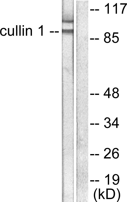
Figure 1. Western blot analysis of Cullin 1 using anti-Cullin 1 antibody (PB9542). Electrophoresis was performed on a 5-20% SDS-PAGE gel at 70V (Stacking gel) / 90V (Resolving gel) for 2-3 hours. Lane 1: Rat Spleen Tissue Lysate at 50ug, Lane 2: Rat Lung Tissue Lysate at 50ug, Lane 3: Rat Thymus Tissue Lysate at 50ug, Lane 4: Mouse Lung Tissue Lysate at 50ug, Lane 5: SW620 Whole Cell Lysate at 40ug. After electrophoresis, proteins were transferred to a nitrocellulose membrane at 150 mA for 50-90 minutes. Blocked the membrane with 5% non-fat milk/TBS for 1.5 hour at RT. The membrane was incubated with rabbit anti-Cullin 1 antigen affinity purified polyclonal antibody (Catalog # PB9542) at 0.5 microg/mL overnight at 4°C, then washed with TBS-0.1%Tween 3 times with 5 minutes each and probed with a goat anti-rabbit IgG-HRP secondary antibody at a dilution of 1:5000 for 1.5 hour at RT. The signal is developed using an Enhanced Chemiluminescent detection (ECL) kit (Catalog # EK1002) with Tanon 5200 system. A specific band was detected for Cullin 1 at approximately 90 kDa. The expected band size for Cullin 1 is at 90 kDa.
Figure 1. Western blot analysis of Cullin 1 using anti-Cullin 1 antibody (PB9542). Electrophoresis was performed on a 5-20% SDS-PAGE gel at 70V (Stacking gel) / 90V (Resolving gel) for 2-3 hours. Lane 1: Rat Spleen Tissue Lysate at 50ug, Lane 2: Rat Lung Tissue Lysate at 50ug, Lane 3: Rat Thymus Tissue Lysate at 50ug, Lane 4: Mouse Lung Tissue Lysate at 50ug, Lane 5: SW620 Whole Cell Lysate at 40ug. After electrophoresis, proteins were transferred to a nitrocellulose membrane at 150 mA for 50-90 minutes. Blocked the membrane with 5% non-fat milk/TBS for 1.5 hour at RT. The membrane was incubated with rabbit anti-Cullin 1 antigen affinity purified polyclonal antibody (Catalog # PB9542) at 0.5 microg/mL overnight at 4°C, then washed with TBS-0.1%Tween 3 times with 5 minutes each and probed with a goat anti-rabbit IgG-HRP secondary antibody at a dilution of 1:5000 for 1.5 hour at RT. The signal is developed using an Enhanced Chemiluminescent detection (ECL) kit (Catalog # EK1002) with Tanon 5200 system. A specific band was detected for Cullin 1 at approximately 90 kDa. The expected band size for Cullin 1 is at 90 kDa.
Anti-Cullin 1/CUL1 Antibody Picoband(r)
PB9542-CARRIER-FREE
ApplicationsWestern Blot
Product group Antibodies
ReactivityBovine, Canine, Equine, Human, Monkey, Mouse, Rabbit, Rat
TargetCUL1
Overview
- SupplierBoster Bio
- Product NameAnti-Cullin 1/CUL1 Antibody Picoband(r)
- Delivery Days Customer9
- Application Supplier NoteTested Species: In-house tested species with positive results. Other applications have not been tested. Optimal dilutions should be determined by end users.
- ApplicationsWestern Blot
- CertificationResearch Use Only
- ClonalityPolyclonal
- Concentration500 ug/ml
- Gene ID8454
- Target nameCUL1
- Target descriptioncullin 1
- Target synonymscullin-1, CUL-1
- HostRabbit
- IsotypeIgG
- Protein IDQ13616
- Protein NameCullin-1
- Scientific DescriptionBoster Bio Anti-Cullin 1/CUL1 Antibody Picoband® catalog # PB9542. Tested in WB applications. This antibody reacts with Human, Mouse, Rat. The brand Picoband indicates this is a premium antibody that guarantees superior quality, high affinity, and strong signals with minimal background in Western blot applications. Only our best-performing antibodies are designated as Picoband, ensuring unmatched performance.
- ReactivityBovine, Canine, Equine, Human, Monkey, Mouse, Rabbit, Rat
- Storage Instruction-20°C,2°C to 8°C
- UNSPSC12352203
Related products
Product group Antibodies
Anti-Cullin 1 AntibodyA95442
ApplicationsImmunoFluorescence, Western Blot, ELISA, ImmunoHistoChemistry
ReactivityHuman, Mouse
- SizePrice
Product group Antibodies
Anti-CUL1 (C-term) Antibody102-22261
ApplicationsWestern Blot
TargetCUL1
- SizePrice
Product group Antibodies
Cullin 1 Recombinant Antibody, AbBy Fluor-594 ConjugatedBSM-61690R-BF594
ApplicationsFlow Cytometry, ImmunoFluorescence, Western Blot
ReactivityHuman, Mouse, Rat
TargetCUL1
- SizePrice
Product group Antibodies
CUL1 AntibodyCSB-PA001830
ApplicationsImmunoFluorescence, Western Blot, ELISA, ImmunoHistoChemistry
ReactivityHuman, Mouse
TargetCUL1
- SizePrice
Product group Antibodies
ApplicationsImmunoPrecipitation, Western Blot, ImmunoCytoChemistry, ImmunoHistoChemistry
ReactivityMouse, Rat
TargetCUL1
- SizePrice
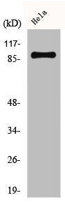
![Various whole cell extracts (30 μg) were separated by 7.5% SDS-PAGE, and the membrane was blotted with Cullin 1 antibody [JM72-30] (GTX00988) diluted at 1:500. The HRP-conjugated anti-rabbit IgG antibody (GTX213110-01) was used to detect the primary antibody.](https://www.genetex.com/upload/website/prouct_img/normal/GTX00988/GTX00988_HK0904_20200214_WB_w_23053121_877.webp)
Product group Antibodies
Cullin 1 antibody [JM72-30]GTX00988
ApplicationsFlow Cytometry, Western Blot, ImmunoHistoChemistry, ImmunoHistoChemistry Paraffin
ReactivityHuman, Mouse, Rat
TargetCUL1
- SizePrice
Product group Antibodies

CUL1 / Cullin 1 AntibodyLS-C331929
ApplicationsImmunoFluorescence, Western Blot, ImmunoHistoChemistry
ReactivityHuman, Mouse, Rat
TargetCUL1
- SizePrice

Product group Antibodies
Anti-CUL1 AntibodyHPA064584
ApplicationsImmunoCytoChemistry
ReactivityHuman
TargetCUL1
- SizePrice
Product group Antibodies
Anti-CUL1Y058318
ApplicationsWestern Blot, ELISA, ImmunoHistoChemistry
ReactivityHuman, Mouse, Rat
- SizePrice