


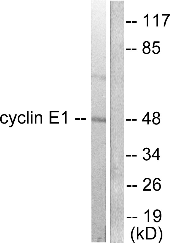
Figure 1. Western blot analysis of CCNE1 using anti-CCNE1 antibody (A00543-2). Electrophoresis was performed on a 5-20% SDS-PAGE gel at 70V (Stacking gel) / 90V (Resolving gel) for 2-3 hours. The sample well of each lane was loaded with 50ug of sample under reducing conditions. Lane 1: human Hela whole cell lysates, Lane 2: human HepG2 whole cell lysates, Lane 3: human K562 whole cell lysates, Lane 4: human U2OS whole cell lysates, Lane 5: human A549 whole cell lysates, Lane 6: human PANC-1 whole cell lysates, Lane 7: human CACO-2 whole cell lysates, After Electrophoresis, proteins were transferred to a Nitrocellulose membrane at 150mA for 50-90 minutes. Blocked the membrane with 5% Non-fat Milk/ TBS for 1.5 hour at RT. The membrane was incubated with rabbit anti-CCNE1 antigen affinity purified polyclonal antibody (Catalog # A00543-2) at 0.25 microg/mL overnight at 4°C, then washed with TBS-0.1%Tween 3 times with 5 minutes each and probed with a goat anti-rabbit IgG-HRP secondary antibody at a dilution of 1:5000 for 1.5 hour at RT. The signal is developed using an Enhanced Chemiluminescent detection (ECL) kit (Catalog # EK1002) with Tanon 5200 system. A specific band was detected for CCNE1 at approximately 47KD. The expected band size for CCNE1 is at 47KD.
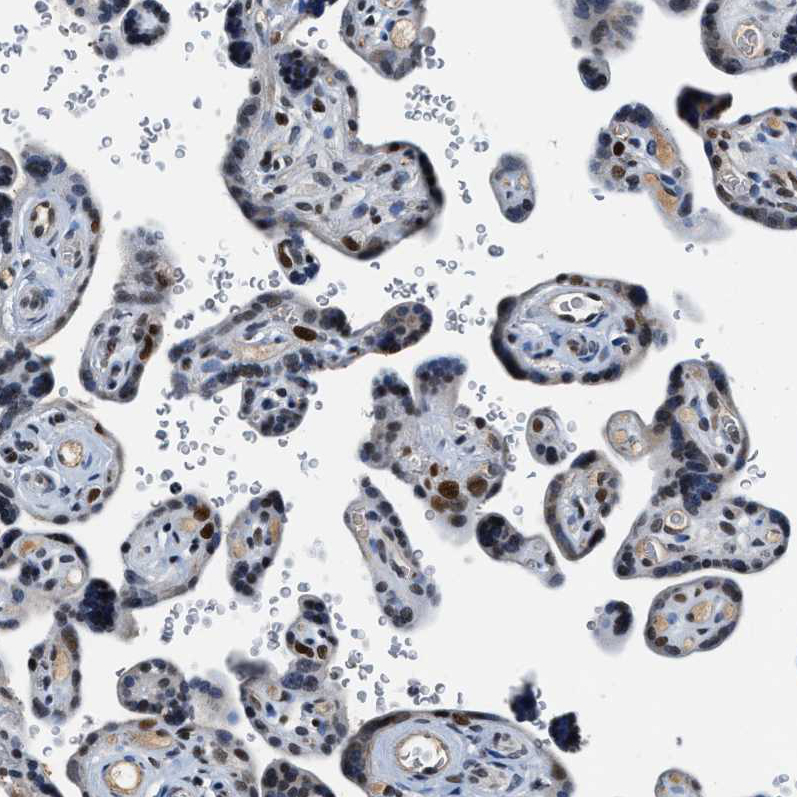
. CCNE1 was detected in paraffin-embedded section of human placenta tissue. Heat mediated antigen retrieval was performed in EDTA buffer (pH8.0, epitope retrieval solution). The tissue section was blocked with 10% goat serum. The tissue section was then incubated with 1microg/ml rabbit anti-CCNE1 Antibody (A00543-2) overnight at 4°C. Biotinylated goat anti-rabbit IgG was used as secondary antibody and incubated for 30 minutes at 37°C. The tissue section was developed using Strepavidin-Biotin-Complex (SABC) (Catalog # SA1022) with DAB as the chromogen.")
. CCNE1 was detected in paraffin-embedded section of human placenta tissue. Heat mediated antigen retrieval was performed in EDTA buffer (pH8.0, epitope retrieval solution). The tissue section was blocked with 10% goat serum. The tissue section was then incubated with 1microg/ml rabbit anti-CCNE1 Antibody (A00543-2) overnight at 4°C. Biotinylated goat anti-rabbit IgG was used as secondary antibody and incubated for 30 minutes at 37°C. The tissue section was developed using Strepavidin-Biotin-Complex (SABC) (Catalog # SA1022) with DAB as the chromogen.")
. CCNE1 was detected in paraffin-embedded section of human lung cancer tissue. Heat mediated antigen retrieval was performed in EDTA buffer (pH8.0, epitope retrieval solution). The tissue section was blocked with 10% goat serum. The tissue section was then incubated with 1microg/ml rabbit anti-CCNE1 Antibody (A00543-2) overnight at 4°C. Biotinylated goat anti-rabbit IgG was used as secondary antibody and incubated for 30 minutes at 37°C. The tissue section was developed using Strepavidin-Biotin-Complex (SABC) (Catalog # SA1022) with DAB as the chromogen.")
. CCNE1 was detected in paraffin-embedded section of mouse testis tissue. Heat mediated antigen retrieval was performed in EDTA buffer (pH8.0, epitope retrieval solution). The tissue section was blocked with 10% goat serum. The tissue section was then incubated with 1microg/ml rabbit anti-CCNE1 Antibody (A00543-2) overnight at 4°C. Biotinylated goat anti-rabbit IgG was used as secondary antibody and incubated for 30 minutes at 37°C. The tissue section was developed using Strepavidin-Biotin-Complex (SABC) (Catalog # SA1022) with DAB as the chromogen.")
. CCNE1 was detected in paraffin-embedded section of rat testis tissue. Heat mediated antigen retrieval was performed in EDTA buffer (pH8.0, epitope retrieval solution). The tissue section was blocked with 10% goat serum. The tissue section was then incubated with 1microg/ml rabbit anti-CCNE1 Antibody (A00543-2) overnight at 4°C. Biotinylated goat anti-rabbit IgG was used as secondary antibody and incubated for 30 minutes at 37°C. The tissue section was developed using Strepavidin-Biotin-Complex (SABC) (Catalog # SA1022) with DAB as the chromogen.")
. Overlay histogram showing SiHa cells stained with A00543-2 (Blue line). To facilitate intracellular staining, cells were fixed with 4% paraformaldehyde and permeabilized with permeabilization buffer. The cells were blocked with 10% normal goat serum. And then incubated with rabbit anti-CCNE1 Antibody (A00543-2, 1microg/1x106 cells) for 30 min at 20°C. DyLight®488 conjugated goat anti-rabbit IgG (BA1127, 5-10microg/1x106 cells) was used as secondary antibody for 30 minutes at 20°C. Isotype control antibody (Green line) was rabbit IgG (1microg/1x106) used under the same conditions. Unlabelled sample without incubation with primary antibody and secondary antibody (Red line) was used as a blank control.")
Figure 1. Western blot analysis of CCNE1 using anti-CCNE1 antibody (A00543-2). Electrophoresis was performed on a 5-20% SDS-PAGE gel at 70V (Stacking gel) / 90V (Resolving gel) for 2-3 hours. The sample well of each lane was loaded with 50ug of sample under reducing conditions. Lane 1: human Hela whole cell lysates, Lane 2: human HepG2 whole cell lysates, Lane 3: human K562 whole cell lysates, Lane 4: human U2OS whole cell lysates, Lane 5: human A549 whole cell lysates, Lane 6: human PANC-1 whole cell lysates, Lane 7: human CACO-2 whole cell lysates, After Electrophoresis, proteins were transferred to a Nitrocellulose membrane at 150mA for 50-90 minutes. Blocked the membrane with 5% Non-fat Milk/ TBS for 1.5 hour at RT. The membrane was incubated with rabbit anti-CCNE1 antigen affinity purified polyclonal antibody (Catalog # A00543-2) at 0.25 microg/mL overnight at 4°C, then washed with TBS-0.1%Tween 3 times with 5 minutes each and probed with a goat anti-rabbit IgG-HRP secondary antibody at a dilution of 1:5000 for 1.5 hour at RT. The signal is developed using an Enhanced Chemiluminescent detection (ECL) kit (Catalog # EK1002) with Tanon 5200 system. A specific band was detected for CCNE1 at approximately 47KD. The expected band size for CCNE1 is at 47KD.
Anti-Cyclin E1/CCNE1 Antibody Picoband(r)
A00543-2-CARRIER-FREE
ApplicationsFlow Cytometry, Western Blot, ELISA, ImmunoHistoChemistry
Product group Antibodies
ReactivityHuman, Mouse, Rat
TargetCCNE1
Overview
- SupplierBoster Bio
- Product NameAnti-Cyclin E1/CCNE1 Antibody Picoband(r)
- Delivery Days Customer9
- ApplicationsFlow Cytometry, Western Blot, ELISA, ImmunoHistoChemistry
- CertificationResearch Use Only
- ClonalityPolyclonal
- Concentration500 ug/ml
- Gene ID898
- Target nameCCNE1
- Target descriptioncyclin E1
- Target synonymsCCNE, pCCNE1, G1/S-specific cyclin-E1
- HostRabbit
- IsotypeIgG
- Protein IDP24864
- Protein NameG1/S-specific cyclin-E1
- Scientific DescriptionBoster Bio Anti-Cyclin E1/CCNE1 Antibody Picoband® catalog # A00543-2. Tested in ELISA, Flow Cytometry, IHC, WB applications. This antibody reacts with Human, Mouse, Rat. The brand Picoband indicates this is a premium antibody that guarantees superior quality, high affinity, and strong signals with minimal background in Western blot applications. Only our best-performing antibodies are designated as Picoband, ensuring unmatched performance.
- ReactivityHuman, Mouse, Rat
- Storage Instruction-20°C,2°C to 8°C
- UNSPSC12352203
Related products
Product group Antibodies
Anti-Cyclin E1 AntibodyA94771
ApplicationsImmunoFluorescence, Western Blot, ELISA, ImmunoHistoChemistry
ReactivityHuman, Mouse, Rat
- SizePrice
Product group Antibodies
Anti-CCNE1 Antibody144-63138
ApplicationsWestern Blot
ReactivityHuman, Mouse, Rat
TargetCCNE1
- SizePrice
Product group Antibodies

CCNE1 / Cyclin E1 AntibodyLS-C831032
ApplicationsELISA, ImmunoHistoChemistry
ReactivityHuman
TargetCCNE1
- SizePrice
![HepG2 cells were stained with Cyclin E1 (4H7) Monoclonal Antibody (bsm-52048R) at [1:200] incubated overnight at 4C, followed by secondary antibody incubation, DAPI staining of the nuclei and detection.](https://biossantibodies.com/image-raw/16228.jpeg)
Product group Antibodies
Cyclin E1 Recombinant AntibodyBSM-52048R
ApplicationsImmunoFluorescence, Western Blot, ImmunoCytoChemistry, ImmunoHistoChemistry, ImmunoHistoChemistry Frozen, ImmunoHistoChemistry Paraffin
ReactivityHuman, Mouse
TargetCCNE1
- SizePrice
Product group Antibodies
CCNE1 AntibodyCSB-PA001879
ApplicationsImmunoFluorescence, Western Blot, ELISA, ImmunoHistoChemistry
ReactivityHuman, Mouse, Rat
TargetCCNE1
- SizePrice
Product group Antibodies
CCNE1 Polyclonal AntibodyCAC14982
ApplicationsWestern Blot, ELISA, ImmunoHistoChemistry
ReactivityMouse
TargetCCNE1
- SizePrice
Product group Antibodies
Cyclin E1 antibodyGTX103045
ApplicationsImmunoFluorescence, Western Blot, ImmunoCytoChemistry
ReactivityHuman, Monkey
TargetCCNE1
- SizePrice
Product group Antibodies
Anti-CCNE1 AntibodyHPA018169
ApplicationsImmunoCytoChemistry, ImmunoHistoChemistry
ReactivityHuman
TargetCCNE1
- SizePrice