

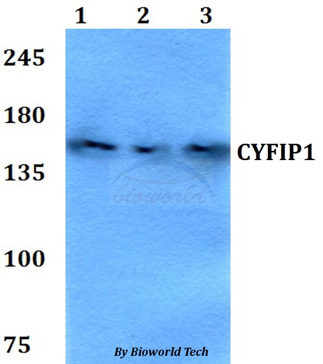
Anti-CYFIP1 Antibody
144-10291
ApplicationsWestern Blot
Product group Antibodies
ReactivityHuman, Mouse, Rat
TargetCYFIP1
Overview
- SupplierRayBiotech
- Product NameAnti-CYFIP1 Antibody
- Delivery Days Customer16
- ApplicationsWestern Blot
- CertificationResearch Use Only
- ClonalityPolyclonal
- ConjugateUnconjugated
- Gene ID23191
- Target nameCYFIP1
- Target descriptioncytoplasmic FMR1 interacting protein 1
- Target synonymsP140SRA-1, SHYC, SRA-1, SRA1, cytoplasmic FMR1-interacting protein 1, cytoplasmic FMRP interacting protein 1, selective hybridizing clone, specifically Rac1-associated protein 1
- HostRabbit
- IsotypeIgG
- Protein IDQ7L576
- Protein NameCytoplasmic FMR1-interacting protein 1
- Scientific DescriptionCYFIP1 Polyclonal Antibody
- ReactivityHuman, Mouse, Rat
- Storage Instruction-20°C
- UNSPSC12352203
Related products
Product group Antibodies
Anti-CYFIP1 AntibodyA28942
ApplicationsWestern Blot
ReactivityHuman, Mouse, Rat
- SizePrice

Product group Antibodies
ApplicationsFlow Cytometry, ImmunoFluorescence, Western Blot, ImmunoCytoChemistry
ReactivityHuman, Mouse, Rat
TargetCYFIP1
- SizePrice

Product group Antibodies
Anti-CYFIP1 AntibodyHPA068106
ApplicationsImmunoHistoChemistry
ReactivityHuman
TargetCYFIP1
- SizePrice
Product group Antibodies
CYFIP1 AntibodyCSB-PA748722LA01HU
ApplicationsImmunoFluorescence, ELISA
ReactivityHuman
TargetCYFIP1
- SizePrice
Product group Antibodies

CYFIP1 AntibodyLS-C496999
ApplicationsWestern Blot
ReactivityHuman, Mouse, Rat
TargetCYFIP1
- SizePrice
Product group Antibodies
CYFIP1 antibody [N1N2], N-termGTX122467
ApplicationsImmunoFluorescence, ImmunoPrecipitation, Western Blot, ImmunoCytoChemistry, ImmunoHistoChemistry, ImmunoHistoChemistry Paraffin
ReactivityHuman, Mouse, Rat
TargetCYFIP1
- SizePrice
Product group Antibodies
ApplicationsFlow Cytometry, Western Blot, ImmunoCytoChemistry
ReactivityHuman, Mouse, Rat
TargetCYFIP1
- SizePrice