

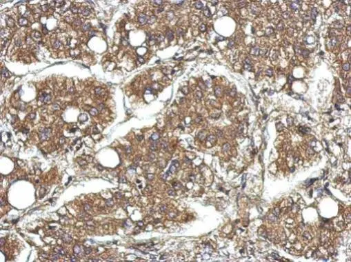
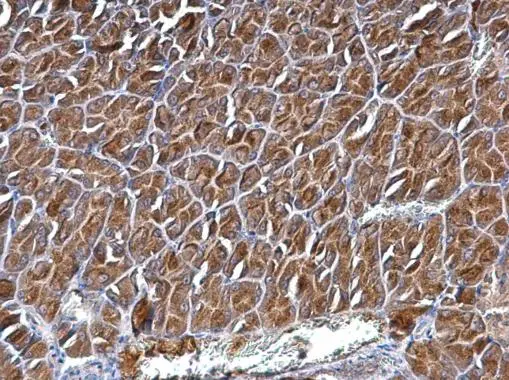
Immunohistochemical analysis of paraffin-embedded human hepatoma, using CYFIP1(GTX122467) antibody at 1:500 dilution.
Antigen Retrieval: Trilogy? (EDTA based, pH 8.0) buffer, 15min
 A: HeLa 5% SDS PAGE GTX122467 diluted at 1:1000 The HRP-conjugated anti-rabbit IgG antibody (GTX213110-01) was used to detect the primary antibody.")
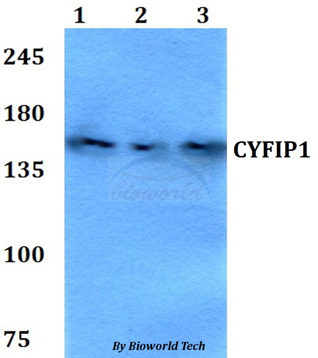
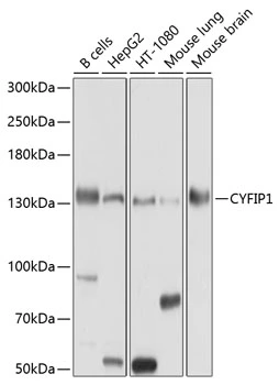
![Various tissue extracts (50 μg) were separated by 5% SDS-PAGE, and the membrane was blotted with CYFIP1 antibody [N1N2], N-term (GTX122467) diluted at 1:1000. The HRP-conjugated anti-rabbit IgG antibody (GTX213110-01) was used to detect the primary antibody.](https://www.genetex.com/upload/website/prouct_img/normal/GTX122467/GTX122467_40828_20170929_WB_M_R_w_23060522_276.webp "Various tissue extracts (50 μg) were separated by 5% SDS-PAGE, and the membrane was blotted with CYFIP1 antibody [N1N2], N-term (GTX122467) diluted at 1:1000. The HRP-conjugated anti-rabbit IgG antibody (GTX213110-01) was used to detect the primary antibody.")
![Immunoprecipitation of CYFIP1 protein from HeLa whole cell extracts using 5 μg of CYFIP1 antibody [N1N2], N-term (GTX122467). Western blot analysis was performed using CYFIP1 antibody [N1N2], N-term (GTX122467). EasyBlot anti-Rabbit IgG (GTX221666-01) was used as a secondary reagent.](https://www.genetex.com/upload/website/prouct_img/normal/GTX122467/GTX122467_40828_20150514_IP_w_23060522_771.webp "Immunoprecipitation of CYFIP1 protein from HeLa whole cell extracts using 5 μg of CYFIP1 antibody [N1N2], N-term (GTX122467). Western blot analysis was performed using CYFIP1 antibody [N1N2], N-term (GTX122467). EasyBlot anti-Rabbit IgG (GTX221666-01) was used as a secondary reagent.")
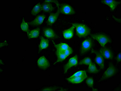
![CYFIP1 antibody [N1N2], N-term detects CYFIP1 protein by immunofluorescent analysis. Sample: DIV9 rat E18 primary cortical neuron cells were fixed in 4% paraformaldehyde at RT for 15 min. Green: CYFIP1 stained by CYFIP1 antibody [N1N2], N-term (GTX122467) diluted at 1:500. Red: beta Tubulin 3/ Tuj1, stained by beta Tubulin 3/ Tuj1 antibody [GT1338] (GTX631831) diluted at 1:500. Blue: Fluoroshield with DAPI (GTX30920).](https://www.genetex.com/upload/website/prouct_img/normal/GTX122467/GTX122467_40828_20171115_ICC_IF_R_w_23060522_185.webp "CYFIP1 antibody [N1N2], N-term detects CYFIP1 protein by immunofluorescent analysis. Sample: DIV9 rat E18 primary cortical neuron cells were fixed in 4% paraformaldehyde at RT for 15 min. Green: CYFIP1 stained by CYFIP1 antibody [N1N2], N-term (GTX122467) diluted at 1:500. Red: beta Tubulin 3/ Tuj1, stained by beta Tubulin 3/ Tuj1 antibody [GT1338] (GTX631831) diluted at 1:500. Blue: Fluoroshield with DAPI (GTX30920).")
Immunohistochemical analysis of paraffin-embedded human hepatoma, using CYFIP1(GTX122467) antibody at 1:500 dilution.
Antigen Retrieval: Trilogy? (EDTA based, pH 8.0) buffer, 15min
CYFIP1 antibody [N1N2], N-term
GTX122467
ApplicationsImmunoFluorescence, ImmunoPrecipitation, Western Blot, ImmunoCytoChemistry, ImmunoHistoChemistry, ImmunoHistoChemistry Paraffin
Product group Antibodies
ReactivityHuman, Mouse, Rat
TargetCYFIP1
Overview
- SupplierGeneTex
- Product NameCYFIP1 antibody [N1N2], N-term
- Delivery Days Customer9
- Application Supplier NoteWB: 1:500-1:3000. ICC/IF: 1:100-1:1000. IHC-P: 1:100-1:1000. IP: 1:100-1:500. *Optimal dilutions/concentrations should be determined by the researcher.Not tested in other applications.
- ApplicationsImmunoFluorescence, ImmunoPrecipitation, Western Blot, ImmunoCytoChemistry, ImmunoHistoChemistry, ImmunoHistoChemistry Paraffin
- CertificationResearch Use Only
- ClonalityPolyclonal
- Concentration1 mg/ml
- ConjugateUnconjugated
- Gene ID23191
- Target nameCYFIP1
- Target descriptioncytoplasmic FMR1 interacting protein 1
- Target synonymsP140SRA-1, SHYC, SRA-1, SRA1, cytoplasmic FMR1-interacting protein 1, cytoplasmic FMRP interacting protein 1, selective hybridizing clone, specifically Rac1-associated protein 1
- HostRabbit
- IsotypeIgG
- Protein IDQ7L576
- Protein NameCytoplasmic FMR1-interacting protein 1
- Scientific DescriptionComponent of the CYFIP1-EIF4E-FMR1 complex which binds to the mRNA cap and mediates translational repression. In the CYFIP1-EIF4E-FMR1 complex this subunit is an adapter between EIF4E and FMR1. Promotes the translation repression activity of FMR1 in brain probably by mediating its association with EIF4E and mRNA (By similarity). Involved in formation of membrane ruffles and lamellipodia protrusions and in axon outgrowth. Binds to F-actin but not to RNA. Regulator of epithelial morphogenesis. May act as an invasion suppressor in cancers.
- ReactivityHuman, Mouse, Rat
- Storage Instruction-20°C or -80°C,2°C to 8°C
- UNSPSC41116161
Datasheet
Related products
Product group Antibodies
Anti-CYFIP1 AntibodyA28942
ApplicationsWestern Blot
ReactivityHuman, Mouse, Rat
- SizePrice

Product group Antibodies
ApplicationsFlow Cytometry, ImmunoFluorescence, Western Blot, ImmunoCytoChemistry
ReactivityHuman, Mouse, Rat
TargetCYFIP1
- SizePrice

Product group Antibodies
Anti-CYFIP1 AntibodyHPA068106
ApplicationsImmunoHistoChemistry
ReactivityHuman
TargetCYFIP1
- SizePrice
Product group Antibodies
CYFIP1 AntibodyCSB-PA748722LA01HU
ApplicationsImmunoFluorescence, ELISA
ReactivityHuman
TargetCYFIP1
- SizePrice
Product group Antibodies

CYFIP1 AntibodyLS-C496999
ApplicationsWestern Blot
ReactivityHuman, Mouse, Rat
TargetCYFIP1
- SizePrice
Product group Antibodies
CYFIP1 antibodyGTX64502
ApplicationsWestern Blot
ReactivityHuman, Mouse
TargetCYFIP1
- SizePrice
Product group Antibodies
CYFIP1 antibodyGTX122802
ApplicationsImmunoHistoChemistry, ImmunoHistoChemistry Paraffin
ReactivityHuman, Mouse
TargetCYFIP1
- SizePrice
Product group Antibodies
ApplicationsFlow Cytometry, Western Blot, ImmunoCytoChemistry
ReactivityHuman, Mouse, Rat
TargetCYFIP1
- SizePrice
Product group Antibodies
Anti-CYFIP1 Antibody144-10291
ApplicationsWestern Blot
ReactivityHuman, Mouse, Rat
TargetCYFIP1
- SizePrice