

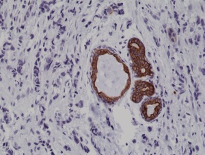
Immunohistochemical staining of formalin fixed and paraffin embedded human breast cancer tissue sections using Anti-CK7 Rabbit Monoclonal Antibody (Clone 284) at a 1:1000 dilution.
Immunohistochemical staining of formalin fixed and paraffin embedded human breast cancer tissue sections using Anti-CK7 Rabbit Monoclonal Antibody (Clone 284) at a 1:1000 dilution.
anti-Cytokeratin-7 (human), Rabbit Monoclonal (RM284)
REV-31-1167-00
ApplicationsWestern Blot, ImmunoHistoChemistry
Product group Antibodies
ReactivityHuman
TargetKRT7
Overview
- SupplierRevMAb Biosciences
- Product Nameanti-Cytokeratin-7 (human), Rabbit Monoclonal (RM284)
- Delivery Days Customer2
- ApplicationsWestern Blot, ImmunoHistoChemistry
- CertificationResearch Use Only
- ClonalityMonoclonal
- Clone IDRM284
- Gene ID3855
- Target nameKRT7
- Target descriptionkeratin 7
- Target synonymsCK7, K2C7, K7, SCL, keratin, type II cytoskeletal 7, CK-7, cytokeratin 7, keratin 7, type II, keratin, 55K type II cytoskeletal, keratin, simple epithelial type I, K7, sarcolectin, type II mesothelial keratin K7, type-II keratin Kb7
- HostRabbit
- IsotypeIgG
- Protein IDP08729
- Protein NameKeratin, type II cytoskeletal 7
- Scientific DescriptionCytokeratins are keratin proteins found in the intracytoplasmic cytoskeleton of epithelial tissue (at least 20 different polypeptides). They are an important component of intermediate filaments, which help cells resist mechanical stress. Expression of these cytokeratins within epithelial cells is largely specific to particular organs or tissues. The subsets of cytokeratins which an epithelial cell expresses depends mainly on the type of epithelium, the moment in the course of terminal differentiation and the stage of development. Thus a specific cytokeratin expression profile allows the identification of epithelial cells. Furthermore, this applies also to the malignant counterparts of the epithelia, (carcinomas). Cytokeratin subtype expression patterns are used to an increasing extent in the distinction of different types of epithelial malignancies. The cytokeratin antibodies are not only of assistance in the differential diagnosis of tumors using immunohistochemistry on tissue sections, but are also a useful tool in cytopathology and flow cytometric assays. Cytokeratin-7 (CK7) is a type II cytokeratin expressed by most ductal, glandular and transitional epithelia. Cytokeratin 7 exhibits a cytoplasmic localization. Because the cytokeratin-7 is found in both healthy and neoplastic cells, antibodies to CK7 can be used in immunohistochemistry to distinguish ovarian and transitional cell carcinomas from colonic and prostate cancers, respectively and is commonly used together with Cytokeratin-20. - Recombinant Antibody. This antibody reacts to human CK7 (Cytokeratin-7). Applications: WB, IHC. Source: Rabbit. Liquid. 50% Glycerol/PBS with 1% BSA and 0.09% sodium azide. Cytokeratins are keratin proteins found in the intracytoplasmic cytoskeleton of epithelial tissue (at least 20 different polypeptides). They are an important component of intermediate filaments, which help cells resist mechanical stress. Expression of these cytokeratins within epithelial cells is largely specific to particular organs or tissues. The subsets of cytokeratins which an epithelial cell expresses depends mainly on the type of epithelium, the moment in the course of terminal differentiation and the stage of development. Thus a specific cytokeratin expression profile allows the identification of epithelial cells. Furthermore, this applies also to the malignant counterparts of the epithelia, (carcinomas). Cytokeratin subtype expression patterns are used to an increasing extent in the distinction of different types of epithelial malignancies. The cytokeratin antibodies are not only of assistance in the differential diagnosis of tumors using immunohistochemistry on tissue sections, but are also a useful tool in cytopathology and flow cytometric assays. Cytokeratin-7 (CK7) is a type II cytokeratin expressed by most ductal, glandular and transitional epithelia. Cytokeratin 7 exhibits a cytoplasmic localization. Because the cytokeratin-7 is found in both healthy and neoplastic cells, antibodies to CK7 can be used in immunohistochemistry to distinguish ovarian and transitional cell carcinomas from colonic and prostate cancers, respectively and is commonly used together with Cytokeratin-20.
- ReactivityHuman
- Storage Instruction-20°C,2°C to 8°C
- UNSPSC41116161
Datasheet
Related products
Product group Antibodies
Anti-Keratin 7 AntibodyA96688
ApplicationsImmunoFluorescence, Western Blot, ELISA, ImmunoHistoChemistry
ReactivityHuman
- SizePrice

Product group Antibodies
Anti-KRT7 Antibody Picoband(r)A02416-2-CARRIER-FREE
ApplicationsWestern Blot, ImmunoHistoChemistry
ReactivityHuman
TargetKRT7
- SizePrice
Product group Antibodies
Anti-Cytokeratin 7 Antibody188-10504
ApplicationsFlow Cytometry
ReactivityHuman
TargetKRT7
- SizePrice
Product group Antibodies

Anti-Cytokeratin 7 [OV-TL 12/30]AB01648-1.1-BT
ApplicationsFlow Cytometry, ImmunoFluorescence, Western Blot, ImmunoHistoChemistry
ReactivityHuman
TargetKRT7
- SizePrice

Product group Antibodies
Anti-KRT7 AntibodyAMAB91530
ApplicationsWestern Blot, ImmunoHistoChemistry
ReactivityHuman
TargetKRT7
- SizePrice
Product group Antibodies
References
CK7 Polyclonal AntibodyBS-1610R
ApplicationsFlow Cytometry, ImmunoFluorescence, Western Blot, ELISA, ImmunoCytoChemistry, ImmunoHistoChemistry, ImmunoHistoChemistry Frozen, ImmunoHistoChemistry Paraffin
ReactivityHuman, Mouse, Rat
TargetKRT7
- SizePrice
Product group Antibodies
KRT7 Monoclonal AntibodyCSB-MA000210
ApplicationsImmunoFluorescence, ImmunoPrecipitation, Western Blot, ELISA, ImmunoHistoChemistry
ReactivityHuman, Mouse, Rat
TargetKRT7
- SizePrice
Product group Antibodies

Cytokeratin 7CK507
ApplicationsImmunoHistoChemistry, ImmunoHistoChemistry Frozen, ImmunoHistoChemistry Paraffin
ReactivityHuman
TargetKRT7
- SizePrice
Product group Antibodies
KRT7 Polyclonal AntibodyCAC14736
ApplicationsImmunoFluorescence, ImmunoPrecipitation, Western Blot, ELISA, ImmunoHistoChemistry
TargetKRT7
- SizePrice