


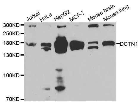
Figure 1. Western blot analysis of DCTN1/p150-glued using anti-DCTN1/p150-glued antibody (A02175-2). Electrophoresis was performed on a 5-20% SDS-PAGE gel at 70V (Stacking gel) / 90V (Resolving gel) for 2-3 hours. The sample well of each lane was loaded with 30ug of sample under reducing conditions. Lane 1: human Hela whole cell lysates, Lane 2: human Jurkat whole cell lysates, Lane 3: human placenta tissue lysates, Lane 4: human U-87MG whole cell lysates, Lane 5: human Caco-2 whole cell lysates, Lane 6: human U20S whole cell lysates, Lane 7: human HEK293 whole cell lysates, Lane 8: human HepG2 whole cell lysates, Lane 9: rat brain tissue lysates, Lane 10: rat stomach tissue lysates, Lane 11: mouse brain tissue lysates. After Electrophoresis, proteins were transferred to a Nitrocellulose membrane at 150mA for 50-90 minutes. Blocked the membrane with 5% Non-fat Milk/ TBS for 1.5 hour at RT. The membrane was incubated with rabbit anti-DCTN1/p150-glued antigen affinity purified polyclonal antibody (Catalog # A02175-2) at 0.25 microg/mL overnight at 4°C, then washed with TBS-0.1%Tween 3 times with 5 minutes each and probed with a goat anti-rabbit IgG-HRP secondary antibody at a dilution of 1:5000 for 1.5 hour at RT. The signal is developed using an Enhanced Chemiluminescent detection (ECL) kit (Catalog # EK1002) with Tanon 5200 system. A specific band was detected for DCTN1/p150-glued at approximately 150KD. The expected band size for DCTN1/p150-glued is at 150KD.
. DCTN1/p150-glued was detected in paraffin-embedded section of human ovarian adenocarcinoma tissue. Heat mediated antigen retrieval was performed in EDTA buffer (pH8.0, epitope retrieval solution). The tissue section was blocked with 10% goat serum. The tissue section was then incubated with 2microg/ml rabbit anti-DCTN1/p150-glued Antibody (A02175-2) overnight at 4°C. Biotinylated goat anti-rabbit IgG was used as secondary antibody and incubated for 30 minutes at 37°C. The tissue section was developed using Strepavidin-Biotin-Complex (SABC) (Catalog # SA1022) with DAB as the chromogen.")
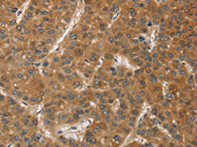
. DCTN1/p150-glued was detected in paraffin-embedded section of human placenta tissue. Heat mediated antigen retrieval was performed in EDTA buffer (pH8.0, epitope retrieval solution). The tissue section was blocked with 10% goat serum. The tissue section was then incubated with 2microg/ml rabbit anti-DCTN1/p150-glued Antibody (A02175-2) overnight at 4°C. Biotinylated goat anti-rabbit IgG was used as secondary antibody and incubated for 30 minutes at 37°C. The tissue section was developed using Strepavidin-Biotin-Complex (SABC) (Catalog # SA1022) with DAB as the chromogen.")
. DCTN1/p150-glued was detected in paraffin-embedded section of human breast cancer tissue. Heat mediated antigen retrieval was performed in EDTA buffer (pH8.0, epitope retrieval solution). The tissue section was blocked with 10% goat serum. The tissue section was then incubated with 2microg/ml rabbit anti-DCTN1/p150-glued Antibody (A02175-2) overnight at 4°C. Biotinylated goat anti-rabbit IgG was used as secondary antibody and incubated for 30 minutes at 37°C. The tissue section was developed using Strepavidin-Biotin-Complex (SABC) (Catalog # SA1022) with DAB as the chromogen.")
. DCTN1/p150-glued was detected in paraffin-embedded section of human breast cancer tissue. Heat mediated antigen retrieval was performed in EDTA buffer (pH8.0, epitope retrieval solution). The tissue section was blocked with 10% goat serum. The tissue section was then incubated with 2microg/ml rabbit anti-DCTN1/p150-glued Antibody (A02175-2) overnight at 4°C. Biotinylated goat anti-rabbit IgG was used as secondary antibody and incubated for 30 minutes at 37°C. The tissue section was developed using Strepavidin-Biotin-Complex (SABC) (Catalog # SA1022) with DAB as the chromogen.")
. DCTN1/p150-glued was detected in paraffin-embedded section of human lung cancer tissue. Heat mediated antigen retrieval was performed in EDTA buffer (pH8.0, epitope retrieval solution). The tissue section was blocked with 10% goat serum. The tissue section was then incubated with 2microg/ml rabbit anti-DCTN1/p150-glued Antibody (A02175-2) overnight at 4°C. Biotinylated goat anti-rabbit IgG was used as secondary antibody and incubated for 30 minutes at 37°C. The tissue section was developed using Strepavidin-Biotin-Complex (SABC) (Catalog # SA1022) with DAB as the chromogen.")
. DCTN1/p150-glued was detected in paraffin-embedded section of human rectal cancer tissue. Heat mediated antigen retrieval was performed in EDTA buffer (pH8.0, epitope retrieval solution). The tissue section was blocked with 10% goat serum. The tissue section was then incubated with 2microg/ml rabbit anti-DCTN1/p150-glued Antibody (A02175-2) overnight at 4°C. Biotinylated goat anti-rabbit IgG was used as secondary antibody and incubated for 30 minutes at 37°C. The tissue section was developed using Strepavidin-Biotin-Complex (SABC) (Catalog # SA1022) with DAB as the chromogen.")
. DCTN1/p150-glued was detected in immunocytochemical section of CACO-2 cells. Enzyme antigen retrieval was performed using IHC enzyme antigen retrieval reagent (AR0022) for 15 mins. The cells were blocked with 10% goat serum. And then incubated with 5microg/mL rabbit anti-DCTN1/p150-glued Antibody (A02175-2) overnight at 4°C. DyLight®488 Conjugated Goat Anti-Rabbit IgG (BA1127) was used as secondary antibody at 1:100 dilution and incubated for 30 minutes at 37°C. The section was counterstained with DAPI. Visualize using a fluorescence microscope and filter sets appropriate for the label used.")
. Overlay histogram showing SiHa cells stained with A02175-2 (Blue line). To facilitate intracellular staining, cells were fixed with 4% paraformaldehyde and permeabilized with permeabilization buffer. The cells were blocked with 10% normal goat serum. And then incubated with rabbit anti-DCTN1/p150-glued Antibody (A02175-2, 1microg/1x106 cells) for 30 min at 20°C. DyLight®488 conjugated goat anti-rabbit IgG (BA1127, 5-10microg/1x106 cells) was used as secondary antibody for 30 minutes at 20°C. Isotype control antibody (Green line) was rabbit IgG (1microg/1x106) used under the same conditions. Unlabelled sample without incubation with primary antibody and secondary antibody (Red line) was used as a blank control.")
Figure 1. Western blot analysis of DCTN1/p150-glued using anti-DCTN1/p150-glued antibody (A02175-2). Electrophoresis was performed on a 5-20% SDS-PAGE gel at 70V (Stacking gel) / 90V (Resolving gel) for 2-3 hours. The sample well of each lane was loaded with 30ug of sample under reducing conditions. Lane 1: human Hela whole cell lysates, Lane 2: human Jurkat whole cell lysates, Lane 3: human placenta tissue lysates, Lane 4: human U-87MG whole cell lysates, Lane 5: human Caco-2 whole cell lysates, Lane 6: human U20S whole cell lysates, Lane 7: human HEK293 whole cell lysates, Lane 8: human HepG2 whole cell lysates, Lane 9: rat brain tissue lysates, Lane 10: rat stomach tissue lysates, Lane 11: mouse brain tissue lysates. After Electrophoresis, proteins were transferred to a Nitrocellulose membrane at 150mA for 50-90 minutes. Blocked the membrane with 5% Non-fat Milk/ TBS for 1.5 hour at RT. The membrane was incubated with rabbit anti-DCTN1/p150-glued antigen affinity purified polyclonal antibody (Catalog # A02175-2) at 0.25 microg/mL overnight at 4°C, then washed with TBS-0.1%Tween 3 times with 5 minutes each and probed with a goat anti-rabbit IgG-HRP secondary antibody at a dilution of 1:5000 for 1.5 hour at RT. The signal is developed using an Enhanced Chemiluminescent detection (ECL) kit (Catalog # EK1002) with Tanon 5200 system. A specific band was detected for DCTN1/p150-glued at approximately 150KD. The expected band size for DCTN1/p150-glued is at 150KD.
Anti-DCTN1/p150-glued Antibody Picoband(r)
A02175-2-CARRIER-FREE
ApplicationsFlow Cytometry, ImmunoFluorescence, Western Blot, ELISA, ImmunoCytoChemistry, ImmunoHistoChemistry
Product group Antibodies
ReactivityHuman, Mouse, Rat
TargetDCTN1
Overview
- SupplierBoster Bio
- Product NameAnti-DCTN1/p150-glued Antibody Picoband(r)
- Delivery Days Customer9
- ApplicationsFlow Cytometry, ImmunoFluorescence, Western Blot, ELISA, ImmunoCytoChemistry, ImmunoHistoChemistry
- CertificationResearch Use Only
- ClonalityPolyclonal
- Concentration500 ug/ml
- Gene ID1639
- Target nameDCTN1
- Target descriptiondynactin subunit 1
- Target synonymsDAP-150, DP-150, HMND14, P135, dynactin subunit 1, 150 kDa dynein-associated polypeptide, dynactin 1 (p150, glued homolog, Drosophila)
- HostRabbit
- IsotypeIgG
- Protein IDQ14203
- Protein NameDynactin subunit 1
- Scientific DescriptionBoster Bio Anti-DCTN1/p150-glued Antibody Picoband® catalog # A02175-2. Tested in ELISA, Flow Cytometry, IF, IHC, ICC, WB applications. This antibody reacts with Human, Mouse, Rat. The brand Picoband indicates this is a premium antibody that guarantees superior quality, high affinity, and strong signals with minimal background in Western blot applications. Only our best-performing antibodies are designated as Picoband, ensuring unmatched performance.
- ReactivityHuman, Mouse, Rat
- Storage Instruction-20°C,2°C to 8°C
- UNSPSC12352203
Related products
Product group Antibodies
Anti-DCTN1 AntibodyA29838
ApplicationsWestern Blot, ImmunoHistoChemistry
ReactivityHuman, Mouse, Rat
- SizePrice
Product group Antibodies
Anti-DCTN1 Antibody144-01783
ApplicationsImmunoFluorescence, ImmunoPrecipitation, Western Blot
ReactivityHuman, Mouse, Rat
TargetDCTN1
- SizePrice
Product group Antibodies

Dynactin AntibodyABX431200
ApplicationsFlow Cytometry, ImmunoFluorescence, Western Blot, ELISA, ImmunoCytoChemistry, ImmunoHistoChemistry
- SizePrice
Product group Antibodies
ApplicationsImmunoFluorescence, Western Blot, ELISA, ImmunoCytoChemistry, ImmunoHistoChemistry, ImmunoHistoChemistry Frozen, ImmunoHistoChemistry Paraffin
ReactivityBovine, Canine, Equine, Human, Mouse, Porcine, Rabbit, Rat
TargetDCTN1
- SizePrice
Product group Antibodies

Goat anti-DynactinEB05639
ApplicationsFlow Cytometry, ImmunoFluorescence, Western Blot, ELISA, ImmunoHistoChemistry
ReactivityHuman, Mouse
TargetDCTN1
- SizePrice
Product group Antibodies
DCTN1 AntibodyCSB-PA038642
ApplicationsWestern Blot, ELISA, ImmunoHistoChemistry
ReactivityHuman, Mouse, Rat
TargetDCTN1
- SizePrice
![Various whole cell extracts (30 μg) were separated by 5% SDS-PAGE, and the membrane was blotted with DCTN1 antibody [C3], C-term (GTX100231) diluted at 1:2500. The HRP-conjugated anti-rabbit IgG antibody (GTX213110-01) was used to detect the primary antibody.](https://www.genetex.com/upload/website/prouct_img/normal/GTX100231/GTX100231_40436_20171124_WB_2_w_23053123_476.webp)
Product group Antibodies
DCTN1 antibody [C3], C-termGTX100231
ApplicationsImmunoFluorescence, Western Blot, ImmunoCytoChemistry, ImmunoHistoChemistry, ImmunoHistoChemistry Paraffin
ReactivityHuman, Mouse, Rat
TargetDCTN1
- SizePrice
Product group Antibodies

DCTN1 / Dynactin 1 AntibodyLS-C335182
ApplicationsImmunoFluorescence, Western Blot, ImmunoHistoChemistry
ReactivityHuman, Mouse, Rat
TargetDCTN1
- SizePrice

Product group Antibodies
Anti-DCTN1 AntibodyHPA069977
ApplicationsImmunoHistoChemistry
ReactivityHuman
TargetDCTN1
- SizePrice