


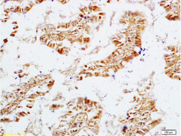
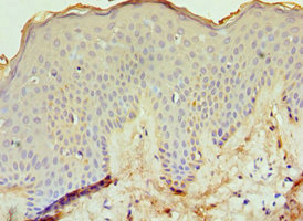
Immunohistochemical staining of human vagina shows strong positivity in endothelial cells.
Immunohistochemical staining of human vagina shows strong positivity in endothelial cells.
Anti-ECE1 Antibody
HPA013616
ApplicationsImmunoHistoChemistry
Product group Antibodies
ReactivityHuman
TargetECE1
Overview
- SupplierAtlas Antibodies
- Product NameAnti-ECE1 Antibody
- Delivery Days Customer4
- ApplicationsImmunoHistoChemistry
- CertificationResearch Use Only
- ClonalityPolyclonal
- ConjugateUnconjugated
- Gene ID1889
- Target nameECE1
- Target descriptionendothelin converting enzyme 1
- Target synonymsECE, endothelin-converting enzyme 1, ECE-1
- HostRabbit
- IsotypeIgG
- Protein IDP42892
- Protein NameEndothelin-converting enzyme 1
- Scientific DescriptionRecombinant Protein Epitope Signature Tag (PrEST) antigen sequence
- ReactivityHuman
- Storage Instruction-20°C,2°C to 8°C
- UNSPSC41116161
Datasheet
MSDS
Related products
Product group Antibodies
Anti-ECE1 AntibodyA30846
ApplicationsWestern Blot, ImmunoHistoChemistry
ReactivityHuman, Mouse, Rat
- SizePrice
Product group Antibodies
Anti-ECE1 Antibody Picoband(r)A02519-1-CARRIER-FREE
ApplicationsWestern Blot
ReactivityHuman, Mouse, Rat
TargetECE1
- SizePrice
Product group Antibodies
Anti-ECE1 Antibody144-05638
ApplicationsWestern Blot
ReactivityHuman, Mouse, Rat
TargetECE1
- SizePrice
Product group Antibodies
ECE1 Polyclonal AntibodyBS-1190R
ApplicationsImmunoFluorescence, ELISA, ImmunoCytoChemistry, ImmunoHistoChemistry, ImmunoHistoChemistry Frozen, ImmunoHistoChemistry Paraffin
ReactivityBovine, Canine, Guinea Pig, Human, Mouse, Porcine, Rabbit, Rat
TargetECE1
- SizePrice
Product group Antibodies
ECE1 AntibodyCSB-PA007371ESR2HU
ApplicationsELISA, ImmunoHistoChemistry
ReactivityHuman
TargetECE1
- SizePrice
Product group Antibodies
ApplicationsImmunoPrecipitation, Western Blot, ImmunoCytoChemistry, ImmunoHistoChemistry
ReactivityPorcine
TargetECE1
- SizePrice
Product group Antibodies

ECE-1 AntibodyLS-C400720
ApplicationsWestern Blot, ELISA
ReactivityHuman, Mouse, Rat
TargetECE1
- SizePrice

Product group Antibodies
Anti-ECE1 AntibodyHPA001490
ApplicationsWestern Blot, ImmunoHistoChemistry
ReactivityHuman
TargetECE1
- SizePrice