


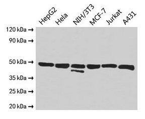
Figure 1. Western blot analysis of ENO1 using anti-ENO1 antibody (A01250-1). Electrophoresis was performed on a 5-20% SDS-PAGE gel at 70V (Stacking gel) / 90V (Resolving gel) for 2-3 hours. The sample well of each lane was loaded with 50ug of sample under reducing conditions. Lane 1: human Hela whole cell lysates, Lane 2: human HepG2 whole cell lysates, Lane 3: human SH-SY5Y whole cell lysates, Lane 4: human U-87MG whole cell lysates, Lane 5: human HEK293 whole cell lysates, Lane 6: human Caco-2 whole cell lysates, Lane 7: monkey kidney tissue lysates, Lane 8: monkey liver tissue lysates. After Electrophoresis, proteins were transferred to a Nitrocellulose membrane at 150mA for 50-90 minutes. Blocked the membrane with 5% Non-fat Milk/ TBS for 1.5 hour at RT. The membrane was incubated with rabbit anti-ENO1 antigen affinity purified polyclonal antibody (Catalog # A01250-1) at 0.25 microg/mL overnight at 4°C, then washed with TBS-0.1%Tween 3 times with 5 minutes each and probed with a goat anti-rabbit IgG-HRP secondary antibody at a dilution of 1:10000 for 1.5 hour at RT. The signal is developed using an Enhanced Chemiluminescent detection (ECL) kit (Catalog # EK1002) with Tanon 5200 system. A specific band was detected for ENO1 at approximately 47KD. The expected band size for ENO1 is at 47KD.
. Electrophoresis was performed on a 5-20% SDS-PAGE gel at 70V (Stacking gel) / 90V (Resolving gel) for 2-3 hours. The sample well of each lane was loaded with 50ug of sample under reducing conditions. Lane 1: rat brain tissue lysates, Lane 2: rat heart tissue lysates, Lane 3: rat kidney tissue lysates, Lane 4: rat liver tissue lysates, Lane 5: mouse brain tissue lysates, Lane 6: mouse kidney tissue lysates, Lane 7: mouse liver tissue lysates, Lane 8: mouse RAW264.7 whole cell lysates. After Electrophoresis, proteins were transferred to a Nitrocellulose membrane at 150mA for 50-90 minutes. Blocked the membrane with 5% Non-fat Milk/ TBS for 1.5 hour at RT. The membrane was incubated with rabbit anti-ENO1 antigen affinity purified polyclonal antibody (Catalog # A01250-1) at 0.25 microg/mL overnight at 4°C, then washed with TBS-0.1%Tween 3 times with 5 minutes each and probed with a goat anti-rabbit IgG-HRP secondary antibody at a dilution of 1:10000 for 1.5 hour at RT. The signal is developed using an Enhanced Chemiluminescent detection (ECL) kit (Catalog # EK1002) with Tanon 5200 system. A specific band was detected for ENO1 at approximately 47KD. The expected band size for ENO1 is at 47KD.")
. ENO1 was detected in paraffin-embedded section of human liver cancer tissue. Heat mediated antigen retrieval was performed in EDTA buffer (pH8.0, epitope retrieval solution). The tissue section was blocked with 10% goat serum. The tissue section was then incubated with 1microg/ml rabbit anti-ENO1 Antibody (A01250-1) overnight at 4°C. Biotinylated goat anti-rabbit IgG was used as secondary antibody and incubated for 30 minutes at 37°C. The tissue section was developed using Strepavidin-Biotin-Complex (SABC) (Catalog # SA1022) with DAB as the chromogen.")
. ENO1 was detected in paraffin-embedded section of mouse testis tissue. Heat mediated antigen retrieval was performed in EDTA buffer (pH8.0, epitope retrieval solution). The tissue section was blocked with 10% goat serum. The tissue section was then incubated with 1microg/ml rabbit anti-ENO1 Antibody (A01250-1) overnight at 4°C. Biotinylated goat anti-rabbit IgG was used as secondary antibody and incubated for 30 minutes at 37°C. The tissue section was developed using Strepavidin-Biotin-Complex (SABC) (Catalog # SA1022) with DAB as the chromogen.")
. ENO1 was detected in paraffin-embedded section of rat testis tissue. Heat mediated antigen retrieval was performed in EDTA buffer (pH8.0, epitope retrieval solution). The tissue section was blocked with 10% goat serum. The tissue section was then incubated with 1microg/ml rabbit anti-ENO1 Antibody (A01250-1) overnight at 4°C. Biotinylated goat anti-rabbit IgG was used as secondary antibody and incubated for 30 minutes at 37°C. The tissue section was developed using Strepavidin-Biotin-Complex (SABC) (Catalog # SA1022) with DAB as the chromogen.")
. ENO1 was detected in immunocytochemical section of A431 cells. Enzyme antigen retrieval was performed using IHC enzyme antigen retrieval reagent (AR0022) for 15 mins. The cells were blocked with 10% goat serum. And then incubated with 2microg/mL rabbit anti-ENO1 Antibody (A01250-1) overnight at 4°C. DyLight®488 Conjugated Goat Anti-Rabbit IgG (BA1127) was used as secondary antibody at 1:100 dilution and incubated for 30 minutes at 37°C. The section was counterstained with DAPI. Visualize using a fluorescence microscope and filter sets appropriate for the label used.")
. Overlay histogram showing HL-60 cells stained with A01250-1 (Blue line). To facilitate intracellular staining, cells were fixed with 4% paraformaldehyde and permeabilized with permeabilization buffer. The cells were blocked with 10% normal goat serum. And then incubated with rabbit anti-ENO1 Antibody (A01250-1,1microg/1x106 cells) for 30 min at 20°C. DyLight®488 conjugated goat anti-rabbit IgG (BA1127, 5-10microg/1x106 cells) was used as secondary antibody for 30 minutes at 20°C. Isotype control antibody (Green line) was rabbit IgG (1microg/1x106) used under the same conditions. Unlabelled sample without incubation with primary antibody and secondary antibody (Red line) was used as a blank control.")
Figure 1. Western blot analysis of ENO1 using anti-ENO1 antibody (A01250-1). Electrophoresis was performed on a 5-20% SDS-PAGE gel at 70V (Stacking gel) / 90V (Resolving gel) for 2-3 hours. The sample well of each lane was loaded with 50ug of sample under reducing conditions. Lane 1: human Hela whole cell lysates, Lane 2: human HepG2 whole cell lysates, Lane 3: human SH-SY5Y whole cell lysates, Lane 4: human U-87MG whole cell lysates, Lane 5: human HEK293 whole cell lysates, Lane 6: human Caco-2 whole cell lysates, Lane 7: monkey kidney tissue lysates, Lane 8: monkey liver tissue lysates. After Electrophoresis, proteins were transferred to a Nitrocellulose membrane at 150mA for 50-90 minutes. Blocked the membrane with 5% Non-fat Milk/ TBS for 1.5 hour at RT. The membrane was incubated with rabbit anti-ENO1 antigen affinity purified polyclonal antibody (Catalog # A01250-1) at 0.25 microg/mL overnight at 4°C, then washed with TBS-0.1%Tween 3 times with 5 minutes each and probed with a goat anti-rabbit IgG-HRP secondary antibody at a dilution of 1:10000 for 1.5 hour at RT. The signal is developed using an Enhanced Chemiluminescent detection (ECL) kit (Catalog # EK1002) with Tanon 5200 system. A specific band was detected for ENO1 at approximately 47KD. The expected band size for ENO1 is at 47KD.
Anti-ENO1 Antibody Picoband(r)
A01250-1-CARRIER-FREE
ApplicationsFlow Cytometry, ImmunoFluorescence, ImmunoPrecipitation, Western Blot, ImmunoCytoChemistry, ImmunoHistoChemistry
Product group Antibodies
ReactivityHuman, Monkey, Mouse, Rat
TargetENO1
Overview
- SupplierBoster Bio
- Product NameAnti-ENO1 Antibody Picoband(r)
- Delivery Days Customer9
- ApplicationsFlow Cytometry, ImmunoFluorescence, ImmunoPrecipitation, Western Blot, ImmunoCytoChemistry, ImmunoHistoChemistry
- CertificationResearch Use Only
- ClonalityPolyclonal
- Concentration500 ug/ml
- Gene ID2023
- Target nameENO1
- Target descriptionenolase 1
- Target synonymsENO1-IT1, ENO1L1, HEL-S-17, MPB1, NNE, PPH, alpha-enolase, c-myc promoter-binding protein-1, 2-phospho-D-glycerate hydro-lyase, ENO1 intronic transcript 1, ENO1 intronic transcript 1 (non-protein coding), MYC promoter-binding protein 1, alpha enolase like 1, enolase 1, (alpha), enolase-alpha, epididymis secretory protein Li 17, non-neural enolase, phosphopyruvate hydratase, plasminogen-binding protein, tau-crystallin
- HostRabbit
- IsotypeIgG
- Protein IDP06733
- Protein NameAlpha-enolase
- Scientific DescriptionBoster Bio Anti-ENO1 Antibody Picoband® catalog # A01250-1. Tested in Flow Cytometry, IP, IF, IHC, ICC, WB applications. This antibody reacts with Human, Monkey, Mouse, Rat. The brand Picoband indicates this is a premium antibody that guarantees superior quality, high affinity, and strong signals with minimal background in Western blot applications. Only our best-performing antibodies are designated as Picoband, ensuring unmatched performance.
- ReactivityHuman, Monkey, Mouse, Rat
- Storage Instruction-20°C,2°C to 8°C
- UNSPSC12352203
Related products
Product group Antibodies

Anti-Alpha-enolase [EnL5]Ab02940-10.0
ApplicationsFlow Cytometry, ImmunoFluorescence, ELISA
ReactivityHuman
TargetENO1
- SizePrice
Product group Antibodies
Anti-ENO1 Antibody144-64672
ApplicationsImmunoFluorescence, Western Blot
ReactivityHuman, Mouse, Rat
TargetENO1
- SizePrice
Product group Antibodies

STRIP1 AntibodyABX126672
ApplicationsImmunoFluorescence, Western Blot, ImmunoCytoChemistry
- SizePrice
Product group Antibodies
ENO1 Recombinant AntibodyBSM-61116R
ApplicationsImmunoFluorescence, ImmunoPrecipitation, Western Blot, ImmunoCytoChemistry
TargetENO1
- SizePrice
Product group Antibodies
Eno1 Polyclonal AntibodyCAC07068
ApplicationsImmunoFluorescence, Western Blot, ELISA, ImmunoHistoChemistry
ReactivityMouse
TargetENO1
- SizePrice
Product group Antibodies
ENO1 AntibodyCSB-PA02395A0RB
ApplicationsImmunoFluorescence, Western Blot, ELISA, ImmunoHistoChemistry
ReactivityHuman, Mouse
TargetENO1
- SizePrice
![Immunohistochemical analysis of paraffin-embedded zebrafish tissue, using ENO1 antibody [N3C3] (GTX101803) at 1:300 dilution.](https://www.genetex.com/upload/website/prouct_img/normal/GTX101803/GTX101803_39799_IHC_Z_22111423_494.webp)
Product group Antibodies
ENO1 antibody [N3C3]GTX101803
ApplicationsDot Blot, ImmunoFluorescence, Western Blot, ImmunoCytoChemistry, ImmunoHistoChemistry, ImmunoHistoChemistry Paraffin
ReactivityHuman, Mouse, Rat, Zebra Fish
TargetENO1
- SizePrice

Product group Antibodies
Anti-ENO1 AntibodyHPA068721
ApplicationsWestern Blot, ImmunoCytoChemistry
ReactivityHuman
TargetENO1
- SizePrice