

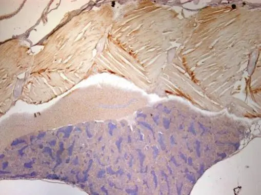
Immunohistochemical analysis of paraffin-embedded zebrafish tissue, using ENO1 antibody [N3C3] (GTX101803) at 1:300 dilution.
![ENO1 antibody [N3C3] detects Eno1 protein on zebrafish by whole mount immunohistochemical analysis. Sample: 2 days-post-fertilization zebrafish embryo. ENO1 antibody [N3C3] (GTX101803) dilution: 1:100.](https://www.genetex.com/upload/website/prouct_img/normal/GTX101803/GTX101803_41738_20160530_IHC-Wm_Z_22111423_194.webp "ENO1 antibody [N3C3] detects Eno1 protein on zebrafish by whole mount immunohistochemical analysis. Sample: 2 days-post-fertilization zebrafish embryo. ENO1 antibody [N3C3] (GTX101803) dilution: 1:100.")
 A: zebrafish eye 10% SDS PAGE GTX101803 diluted at 1:1000")
![ENO1 antibody [N3C3] detects ENO1 protein at cytoplasm by immunofluorescent analysis. Sample: HeLa cells were fixed in 4% paraformaldehyde at RT for 15 min. Green: ENO1 protein stained by ENO1 antibody [N3C3] (GTX101803) diluted at 1:200. Red: alpha Tubulin, a cytoskeleton marker, stained by alpha Tubulin antibody [B-5-1-2] (GTX11304) diluted at 1:10000. Blue: Hoechst 33342 staining.](https://www.genetex.com/upload/website/prouct_img/normal/GTX101803/GTX101803_39799_20150410_IFA_w_23060100_918.webp "ENO1 antibody [N3C3] detects ENO1 protein at cytoplasm by immunofluorescent analysis. Sample: HeLa cells were fixed in 4% paraformaldehyde at RT for 15 min. Green: ENO1 protein stained by ENO1 antibody [N3C3] (GTX101803) diluted at 1:200. Red: alpha Tubulin, a cytoskeleton marker, stained by alpha Tubulin antibody [B-5-1-2] (GTX11304) diluted at 1:10000. Blue: Hoechst 33342 staining.")
 A: Mouse brain 10% SDS PAGE GTX101803 diluted at 1:1000 The HRP-conjugated anti-rabbit IgG antibody (GTX213110-01) was used to detect the primary antibody.")
![Rat tissue extract (50 μg) was separated by 10% SDS-PAGE, and the membrane was blotted with ENO1 antibody [N3C3] (GTX101803) diluted at 1:1000. The HRP-conjugated anti-rabbit IgG antibody (GTX213110-01) was used to detect the primary antibody, and the signal was developed with Trident ECL plus-Enhanced.](https://www.genetex.com/upload/website/prouct_img/normal/GTX101803/GTX101803_39799_20200424_WB_R_lung_w_23060100_281.webp "Rat tissue extract (50 μg) was separated by 10% SDS-PAGE, and the membrane was blotted with ENO1 antibody [N3C3] (GTX101803) diluted at 1:1000. The HRP-conjugated anti-rabbit IgG antibody (GTX213110-01) was used to detect the primary antibody, and the signal was developed with Trident ECL plus-Enhanced.")
 A: A431 (GTX27909) B: H1299 10% SDS PAGE GTX101803 diluted at 1:3000 The HRP-conjugated anti-rabbit IgG antibody (GTX213110-01) was used to detect the primary antibody.")
 antibody at 1:100 dilution.
Antigen Retrieval: Trilogy? (EDTA based, pH 8.0) buffer, 15min")
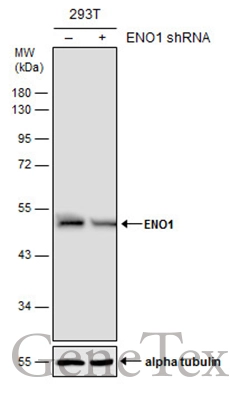
![Non-transfected (–) and transfected (+) 293T whole cell extracts (30 μg) were separated by 10% SDS-PAGE, and the membrane was blotted with ENO1 antibody [N3C3] (GTX101803) diluted at 1:20000. The HRP-conjugated anti-rabbit IgG antibody (GTX213110-01) was used to detect the primary antibody.](https://www.genetex.com/upload/website/prouct_img/normal/GTX101803/GTX101803_39799_20161103_WB_shRNA_watermark_w_23060100_296.webp "Non-transfected (–) and transfected (+) 293T whole cell extracts (30 μg) were separated by 10% SDS-PAGE, and the membrane was blotted with ENO1 antibody [N3C3] (GTX101803) diluted at 1:20000. The HRP-conjugated anti-rabbit IgG antibody (GTX213110-01) was used to detect the primary antibody.")
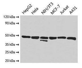
![Various whole cell extracts (30 μg) were separated by 10% SDS-PAGE, and the membrane was blotted with ENO1 antibody [N3C3] (GTX101803) diluted at 1:1000. The HRP-conjugated anti-rabbit IgG antibody (GTX213110-01) was used to detect the primary antibody.](https://www.genetex.com/upload/website/prouct_img/normal/GTX101803/GTX101803_39799_20200424_WB_R_w_23060100_658.webp "Various whole cell extracts (30 μg) were separated by 10% SDS-PAGE, and the membrane was blotted with ENO1 antibody [N3C3] (GTX101803) diluted at 1:1000. The HRP-conjugated anti-rabbit IgG antibody (GTX213110-01) was used to detect the primary antibody.")
Immunohistochemical analysis of paraffin-embedded zebrafish tissue, using ENO1 antibody [N3C3] (GTX101803) at 1:300 dilution.
ENO1 antibody [N3C3]
GTX101803
ApplicationsDot Blot, ImmunoFluorescence, Western Blot, ImmunoCytoChemistry, ImmunoHistoChemistry, ImmunoHistoChemistry Paraffin
Product group Antibodies
ReactivityHuman, Mouse, Rat, Zebra Fish
TargetENO1
Overview
- SupplierGeneTex
- Product NameENO1 antibody [N3C3]
- Delivery Days Customer9
- Application Supplier NoteWB: 1:500-1:20000. ICC/IF: 1:100-1:1000. IHC-P: 1:100-1:1000. *Optimal dilutions/concentrations should be determined by the researcher.Not tested in other applications.
- ApplicationsDot Blot, ImmunoFluorescence, Western Blot, ImmunoCytoChemistry, ImmunoHistoChemistry, ImmunoHistoChemistry Paraffin
- CertificationResearch Use Only
- ClonalityPolyclonal
- Concentration1 mg/ml
- ConjugateUnconjugated
- Gene ID2023
- Target nameENO1
- Target descriptionenolase 1
- Target synonymsENO1-IT1, ENO1L1, HEL-S-17, MPB1, NNE, PPH, alpha-enolase, c-myc promoter-binding protein-1, 2-phospho-D-glycerate hydro-lyase, ENO1 intronic transcript 1, ENO1 intronic transcript 1 (non-protein coding), MYC promoter-binding protein 1, alpha enolase like 1, enolase 1, (alpha), enolase-alpha, epididymis secretory protein Li 17, non-neural enolase, phosphopyruvate hydratase, plasminogen-binding protein, tau-crystallin
- HostRabbit
- IsotypeIgG
- Protein IDP06733
- Protein NameAlpha-enolase
- Scientific DescriptionThis gene encodes one of three enolase isoenzymes found in mammals; it encodes alpha-enolase, a homodimeric soluble enzyme, and also encodes a shorter monomeric structural lens protein, tau-crystallin. The two proteins are made from the same message. The full length protein, the isoenzyme, is found in the cytoplasm. The shorter protein is produced from an alternative translation start, is localized to the nucleus, and has been found to bind to an element in the c-myc promoter. A pseudogene has been identified that is located on the other arm of the same chromosome. [provided by RefSeq]
- ReactivityHuman, Mouse, Rat, Zebra Fish
- Storage Instruction-20°C or -80°C,2°C to 8°C
- UNSPSC41116161
Datasheet
Related products
Product group Antibodies

Anti-Alpha-enolase [EnL5]Ab02940-10.0
ApplicationsFlow Cytometry, ImmunoFluorescence, ELISA
ReactivityHuman
TargetENO1
- SizePrice

Product group Antibodies
Anti-ENO1 Antibody Picoband(r)A01250-1-CARRIER-FREE
ApplicationsFlow Cytometry, ImmunoFluorescence, ImmunoPrecipitation, Western Blot, ImmunoCytoChemistry, ImmunoHistoChemistry
ReactivityHuman, Monkey, Mouse, Rat
TargetENO1
- SizePrice
Product group Antibodies
Anti-ENO1 Antibody144-64672
ApplicationsImmunoFluorescence, Western Blot
ReactivityHuman, Mouse, Rat
TargetENO1
- SizePrice
Product group Antibodies

STRIP1 AntibodyABX126672
ApplicationsImmunoFluorescence, Western Blot, ImmunoCytoChemistry
- SizePrice
Product group Antibodies
ENO1 Recombinant AntibodyBSM-61116R
ApplicationsImmunoFluorescence, ImmunoPrecipitation, Western Blot, ImmunoCytoChemistry
TargetENO1
- SizePrice
Product group Antibodies
Eno1 Polyclonal AntibodyCAC07068
ApplicationsImmunoFluorescence, Western Blot, ELISA, ImmunoHistoChemistry
ReactivityMouse
TargetENO1
- SizePrice
Product group Antibodies
ENO1 AntibodyCSB-PA02395A0RB
ApplicationsImmunoFluorescence, Western Blot, ELISA, ImmunoHistoChemistry
ReactivityHuman, Mouse
TargetENO1
- SizePrice

Product group Antibodies
Anti-ENO1 AntibodyHPA068721
ApplicationsWestern Blot, ImmunoCytoChemistry
ReactivityHuman
TargetENO1
- SizePrice
Product group Antibodies
ENO1 antibodyGTX113179
ApplicationsImmunoFluorescence, ImmunoPrecipitation, Western Blot, ImmunoCytoChemistry, ImmunoHistoChemistry, ImmunoHistoChemistry Paraffin
ReactivityHuman, Mouse, Rat
TargetENO1
- SizePrice