


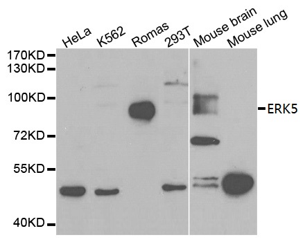
Figure 1. Western blot analysis of ERK5/MAPK7 using anti-ERK5/MAPK7 antibody (A02812-3). Electrophoresis was performed on a 5-20% SDS-PAGE gel at 70V (Stacking gel) / 90V (Resolving gel) for 2-3 hours. The sample well of each lane was loaded with 50ug of sample under reducing conditions. Lane 1: human K562 whole cell lysates, Lane 2: human Hek293 whole cell lysates, Lane 3: human Hela whole cell lysates, Lane 4: monkey Cos-7 whole cell lysates, Lane 5: rat brain tissue lysates, Lane 6: mouse brain tissue lysates, Lane 7: mouse NIH/3T3 whole cell lysates. After Electrophoresis, proteins were transferred to a Nitrocellulose membrane at 150mA for 50-90 minutes. Blocked the membrane with 5% Non-fat Milk/ TBS for 1.5 hour at RT. The membrane was incubated with rabbit anti-ERK5/MAPK7 antigen affinity purified polyclonal antibody (Catalog # A02812-3) at 0.5 microg/mL overnight at 4°C, then washed with TBS-0.1%Tween 3 times with 5 minutes each and probed with a goat anti-rabbit IgG-HRP secondary antibody at a dilution of 1:5000 for 1.5 hour at RT. The signal is developed using an Enhanced Chemiluminescent detection (ECL) kit (Catalog # EK1002) with Tanon 5200 system. A specific band was detected for ERK5/MAPK7 at approximately 115-120KD. The expected band size for ERK5/MAPK7 is at 88KD.
. ERK5/MAPK7 was detected in paraffin-embedded section of human breast cancer tissue. Heat mediated antigen retrieval was performed in EDTA buffer (pH8.0, epitope retrieval solution). The tissue section was blocked with 10% goat serum. The tissue section was then incubated with 2microg/ml rabbit anti-ERK5/MAPK7 Antibody (A02812-3) overnight at 4°C. Biotinylated goat anti-rabbit IgG was used as secondary antibody and incubated for 30 minutes at 37°C. The tissue section was developed using Strepavidin-Biotin-Complex (SABC) (Catalog # SA1022) with DAB as the chromogen.")
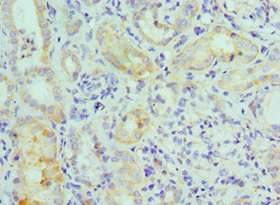
. ERK5/MAPK7 was detected in paraffin-embedded section of human lung cancer tissue. Heat mediated antigen retrieval was performed in EDTA buffer (pH8.0, epitope retrieval solution). The tissue section was blocked with 10% goat serum. The tissue section was then incubated with 2microg/ml rabbit anti-ERK5/MAPK7 Antibody (A02812-3) overnight at 4°C. Biotinylated goat anti-rabbit IgG was used as secondary antibody and incubated for 30 minutes at 37°C. The tissue section was developed using Strepavidin-Biotin-Complex (SABC) (Catalog # SA1022) with DAB as the chromogen.")
Figure 1. Western blot analysis of ERK5/MAPK7 using anti-ERK5/MAPK7 antibody (A02812-3). Electrophoresis was performed on a 5-20% SDS-PAGE gel at 70V (Stacking gel) / 90V (Resolving gel) for 2-3 hours. The sample well of each lane was loaded with 50ug of sample under reducing conditions. Lane 1: human K562 whole cell lysates, Lane 2: human Hek293 whole cell lysates, Lane 3: human Hela whole cell lysates, Lane 4: monkey Cos-7 whole cell lysates, Lane 5: rat brain tissue lysates, Lane 6: mouse brain tissue lysates, Lane 7: mouse NIH/3T3 whole cell lysates. After Electrophoresis, proteins were transferred to a Nitrocellulose membrane at 150mA for 50-90 minutes. Blocked the membrane with 5% Non-fat Milk/ TBS for 1.5 hour at RT. The membrane was incubated with rabbit anti-ERK5/MAPK7 antigen affinity purified polyclonal antibody (Catalog # A02812-3) at 0.5 microg/mL overnight at 4°C, then washed with TBS-0.1%Tween 3 times with 5 minutes each and probed with a goat anti-rabbit IgG-HRP secondary antibody at a dilution of 1:5000 for 1.5 hour at RT. The signal is developed using an Enhanced Chemiluminescent detection (ECL) kit (Catalog # EK1002) with Tanon 5200 system. A specific band was detected for ERK5/MAPK7 at approximately 115-120KD. The expected band size for ERK5/MAPK7 is at 88KD.
Anti-ERK5/MAPK7 Antibody Picoband(r)
A02812-3-CARRIER-FREE
ApplicationsWestern Blot, ELISA, ImmunoHistoChemistry
Product group Antibodies
ReactivityHuman, Monkey, Mouse, Rat
TargetMAPK7
Overview
- SupplierBoster Bio
- Product NameAnti-ERK5/MAPK7 Antibody Picoband(r)
- Delivery Days Customer9
- ApplicationsWestern Blot, ELISA, ImmunoHistoChemistry
- CertificationResearch Use Only
- ClonalityPolyclonal
- Concentration500 ug/ml
- Gene ID5598
- Target nameMAPK7
- Target descriptionmitogen-activated protein kinase 7
- Target synonymsBMK1, ERK4, ERK5, PRKM7, mitogen-activated protein kinase 7, BMK-1, BMK1 kinase, ERK-5, MAP kinase 7, MAPK 7, big MAP kinase 1, extracellular-signal-regulated kinase 5
- HostRabbit
- IsotypeIgG
- Protein IDQ13164
- Protein NameMitogen-activated protein kinase 7
- Scientific DescriptionBoster Bio Anti-ERK5/MAPK7 Antibody Picoband® catalog # A02812-3. Tested in ELISA, IHC, WB applications. This antibody reacts with Human, Monkey, Mouse, Rat. The brand Picoband indicates this is a premium antibody that guarantees superior quality, high affinity, and strong signals with minimal background in Western blot applications. Only our best-performing antibodies are designated as Picoband, ensuring unmatched performance.
- ReactivityHuman, Monkey, Mouse, Rat
- Storage Instruction-20°C,2°C to 8°C
- UNSPSC12352203
Related products
Product group Antibodies
Anti-ERK5 AntibodyA29958
ApplicationsImmunoFluorescence, Western Blot, ImmunoHistoChemistry
ReactivityHuman, Mouse, Rat
- SizePrice
Product group Antibodies
Anti-MAPK7 Antibody144-02111
ApplicationsImmunoFluorescence, Western Blot, ImmunoHistoChemistry
ReactivityHuman, Mouse, Rat
TargetMAPK7
- SizePrice
Product group Antibodies
References
ApplicationsImmunoFluorescence, Western Blot, ELISA, ImmunoCytoChemistry, ImmunoHistoChemistry, ImmunoHistoChemistry Frozen, ImmunoHistoChemistry Paraffin
ReactivityHuman, Mouse, Rat
TargetMAPK7
- SizePrice
Product group Antibodies
MAPK7 AntibodyCSB-PA013465ESR1HU
ApplicationsImmunoFluorescence, ELISA, ImmunoHistoChemistry
ReactivityHuman
TargetMAPK7
- SizePrice
Product group Antibodies
ApplicationsImmunoPrecipitation, Western Blot, ImmunoCytoChemistry, ImmunoHistoChemistry
ReactivityMouse, Porcine, Rat
TargetMAPK7
- SizePrice
Product group Antibodies

MAPK7 / ERK5 AntibodyLS-C403981
ApplicationsWestern Blot, ELISA, ImmunoHistoChemistry
ReactivityHuman, Mouse, Rat
TargetMAPK7
- SizePrice
![Various tissue extracts (50 μg) were separated by 7.5% SDS-PAGE, and the membrane was blotted with ERK5 antibody [GT1258] (GTX02855) diluted at 1:500. The HRP-conjugated anti-rabbit IgG antibody (GTX213110-01) was used to detect the primary antibody.](https://www.genetex.com/upload/website/prouct_img/normal/GTX02855/GTX02855_4000000871_20210122_WB_M_R_w_23053123_881.webp)
Product group Antibodies
ERK5 antibody [GT1258]GTX02855
ApplicationsWestern Blot, ImmunoHistoChemistry, ImmunoHistoChemistry Paraffin
ReactivityHuman, Mouse, Rat
TargetMAPK7
- SizePrice

Product group Antibodies
Anti-MAPK7 AntibodyHPA031031
ApplicationsImmunoCytoChemistry
ReactivityHuman
TargetMAPK7
- SizePrice
Product group Antibodies
Anti-ERK5 AntibodyCAB2111
ApplicationsImmunoFluorescence, Western Blot, ELISA, ImmunoCytoChemistry, ImmunoHistoChemistry, ImmunoHistoChemistry Paraffin
ReactivityHuman
TargetMAPK7
- SizePrice