


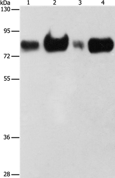
Figure 1. Western blot analysis of FACL4/ACSL4 using anti-FACL4/ACSL4 antibody (M04372). Electrophoresis was performed on a 5-20% SDS-PAGE gel at 70V (Stacking gel) / 90V (Resolving gel) for 2-3 hours. The sample well of each lane was loaded with 30 ug of sample under reducing conditions. Lane 1: human HepG2 whole cell lysates, Lane 2: human PC-3 whole cell lysates, Lane 3: human Hela whole cell lysates, Lane 4: human Caco-2 whole cell lysates. After electrophoresis, proteins were transferred to a nitrocellulose membrane at 150 mA for 50-90 minutes. Blocked the membrane with 5% non-fat milk/TBS for 1.5 hour at RT. The membrane was incubated with mouse anti-FACL4/ACSL4 antigen affinity purified monoclonal antibody (Catalog # M04372) at 0.5 microg/mL overnight at 4°C, then washed with TBS-0.1%Tween 3 times with 5 minutes each and probed with a goat anti-mouse IgG-HRP secondary antibody at a dilution of 1:10000 for 1.5 hour at RT. The signal is developed using an Enhanced Chemiluminescent detection (ECL) kit (Catalog # EK1001) with Tanon 5200 system. A specific band was detected for FACL4/ACSL4 at approximately 79 kDa. The expected band size for FACL4/ACSL4 is at 68 kDa.
. FACL4/ACSL4 was detected in a paraffin-embedded section of human bladder epithelial carcinoma tissue. Heat mediated antigen retrieval was performed in EDTA buffer (pH 8.0, epitope retrieval solution). The tissue section was blocked with 10% goat serum. The tissue section was then incubated with 2 microg/ml mouse anti-FACL4/ACSL4 Antibody (M04372) overnight at 4°C. Biotinylated goat anti-mouse IgG was used as secondary antibody and incubated for 30 minutes at 37°C. The tissue section was developed using Strepavidin-Biotin-Complex (SABC) (Catalog # SA1021) with DAB as the chromogen.")
. FACL4/ACSL4 was detected in a paraffin-embedded section of human lung cancer tissue. Heat mediated antigen retrieval was performed in EDTA buffer (pH 8.0, epitope retrieval solution). The tissue section was blocked with 10% goat serum. The tissue section was then incubated with 2 microg/ml mouse anti-FACL4/ACSL4 Antibody (M04372) overnight at 4°C. Biotinylated goat anti-mouse IgG was used as secondary antibody and incubated for 30 minutes at 37°C. The tissue section was developed using Strepavidin-Biotin-Complex (SABC) (Catalog # SA1021) with DAB as the chromogen.")
. FACL4/ACSL4 was detected in a paraffin-embedded section of human metaplasia of squamous cells of the renal pelvis tissue. Heat mediated antigen retrieval was performed in EDTA buffer (pH 8.0, epitope retrieval solution). The tissue section was blocked with 10% goat serum. The tissue section was then incubated with 2 microg/ml mouse anti-FACL4/ACSL4 Antibody (M04372) overnight at 4°C. Biotinylated goat anti-mouse IgG was used as secondary antibody and incubated for 30 minutes at 37°C. The tissue section was developed using Strepavidin-Biotin-Complex (SABC) (Catalog # SA1021) with DAB as the chromogen.")
. FACL4/ACSL4 was detected in a paraffin-embedded section of human ovarian cancer tissue. Heat mediated antigen retrieval was performed in EDTA buffer (pH 8.0, epitope retrieval solution). The tissue section was blocked with 10% goat serum. The tissue section was then incubated with 2 microg/ml mouse anti-FACL4/ACSL4 Antibody (M04372) overnight at 4°C. Biotinylated goat anti-mouse IgG was used as secondary antibody and incubated for 30 minutes at 37°C. The tissue section was developed using Strepavidin-Biotin-Complex (SABC) (Catalog # SA1021) with DAB as the chromogen.")
. FACL4/ACSL4 was detected in a paraffin-embedded section of human rectal moderately differentiated adenocarcinoma tissue. Heat mediated antigen retrieval was performed in EDTA buffer (pH 8.0, epitope retrieval solution). The tissue section was blocked with 10% goat serum. The tissue section was then incubated with 2 microg/ml mouse anti-FACL4/ACSL4 Antibody (M04372) overnight at 4°C. Biotinylated goat anti-mouse IgG was used as secondary antibody and incubated for 30 minutes at 37°C. The tissue section was developed using Strepavidin-Biotin-Complex (SABC) (Catalog # SA1021) with DAB as the chromogen.")
. FACL4/ACSL4 was detected in an immunocytochemical section of SiHa cells. Enzyme antigen retrieval was performed using IHC enzyme antigen retrieval reagent (AR0022) for 15 mins. The cells were blocked with 10% goat serum. And then incubated with 5 microg/mL mouse anti-FACL4/ACSL4 Antibody (M04372) overnight at 4°C. DyLight®488 Conjugated Goat Anti-Mouse IgG (BA1126) was used as secondary antibody at 1:100 dilution and incubated for 30 minutes at 37°C. The section was counterstained with DAPI. Visualize using a fluorescence microscope and filter sets appropriate for the label used.")
. Overlay histogram showing HepG2 cells stained with M04372 (Blue line). To facilitate intracellular staining, cells were fixed with 4% paraformaldehyde and permeabilized with permeabilization buffer. The cells were blocked with 10% normal goat serum. And then incubated with mouse anti-FACL4/ACSL4 Antibody (M04372, 1 microg/1x106 cells) for 30 min at 20°C. DyLight®488 conjugated goat anti-mouse IgG (BA1126, 5-10 microg/1x106 cells) was used as secondary antibody for 30 minutes at 20°C. Isotype control antibody (Green line) was mouse IgG (1 microg/1x106) used under the same conditions. Unlabelled sample without incubation with primary antibody and secondary antibody (Red line) was used as a blank control.")
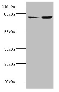
Figure 1. Western blot analysis of FACL4/ACSL4 using anti-FACL4/ACSL4 antibody (M04372). Electrophoresis was performed on a 5-20% SDS-PAGE gel at 70V (Stacking gel) / 90V (Resolving gel) for 2-3 hours. The sample well of each lane was loaded with 30 ug of sample under reducing conditions. Lane 1: human HepG2 whole cell lysates, Lane 2: human PC-3 whole cell lysates, Lane 3: human Hela whole cell lysates, Lane 4: human Caco-2 whole cell lysates. After electrophoresis, proteins were transferred to a nitrocellulose membrane at 150 mA for 50-90 minutes. Blocked the membrane with 5% non-fat milk/TBS for 1.5 hour at RT. The membrane was incubated with mouse anti-FACL4/ACSL4 antigen affinity purified monoclonal antibody (Catalog # M04372) at 0.5 microg/mL overnight at 4°C, then washed with TBS-0.1%Tween 3 times with 5 minutes each and probed with a goat anti-mouse IgG-HRP secondary antibody at a dilution of 1:10000 for 1.5 hour at RT. The signal is developed using an Enhanced Chemiluminescent detection (ECL) kit (Catalog # EK1001) with Tanon 5200 system. A specific band was detected for FACL4/ACSL4 at approximately 79 kDa. The expected band size for FACL4/ACSL4 is at 68 kDa.
Anti-FACL4/ACSL4 Antibody Picoband(r) (monoclonal, 4I7)
M04372-DYLIGHT488
ApplicationsFlow Cytometry, ImmunoFluorescence, Western Blot, ImmunoCytoChemistry, ImmunoHistoChemistry
Product group Antibodies
ReactivityHuman
TargetACSL4
Overview
- SupplierBoster Bio
- Product NameAnti-FACL4/ACSL4 Antibody Picoband(r) (monoclonal, 4I7)
- Delivery Days Customer9
- Application Supplier NoteTested Species: In-house tested species with positive results. Other applications have not been tested. Optimal dilutions should be determined by end users.
- ApplicationsFlow Cytometry, ImmunoFluorescence, Western Blot, ImmunoCytoChemistry, ImmunoHistoChemistry
- CertificationResearch Use Only
- ClonalityMonoclonal
- Clone ID4I7
- Concentration500 ug/ml
- ConjugateDyLight 488
- Gene ID2182
- Target nameACSL4
- Target descriptionacyl-CoA synthetase long chain family member 4
- Target synonymsACS4, FACL4, LACS4, MRX63, MRX68, XLID63, long-chain-fatty-acid--CoA ligase 4, acyl-CoA synthetase 4, arachidonate--CoA ligase, fatty-acid-Coenzyme A ligase, long-chain 4, lignoceroyl-CoA synthase, long-chain acyl-CoA synthetase 4, long-chain fatty-acid-Coenzyme A ligase 4
- HostMouse
- IsotypeIgG1
- Protein IDO60488
- Protein NameLong-chain-fatty-acid--CoA ligase 4
- Scientific DescriptionBoster Bio Anti-FACL4/ACSL4 Antibody Picoband® (monoclonal, 4I7) catalog # M04372. Tested in Flow Cytometry, IF, IHC, ICC, WB applications. This antibody reacts with Human. The brand Picoband indicates this is a premium antibody that guarantees superior quality, high affinity, and strong signals with minimal background in Western blot applications. Only our best-performing antibodies are designated as Picoband, ensuring unmatched performance.
- ReactivityHuman
- Storage Instruction-20°C,2°C to 8°C
- UNSPSC12352203
Related products
Product group Antibodies
Anti-ACSL4 Antibody144-06826
ApplicationsWestern Blot
ReactivityHuman, Mouse
TargetACSL4
- SizePrice
Product group Antibodies
Acsl4 Polyclonal AntibodyCAC09075
ApplicationsWestern Blot, ELISA, ImmunoHistoChemistry
TargetACSL4
- SizePrice
Product group Antibodies
ACSL4 Recombinant AntibodyBSM-62432R
ApplicationsFlow Cytometry, ImmunoFluorescence, ImmunoPrecipitation, Western Blot, ImmunoCytoChemistry, ImmunoHistoChemistry, ImmunoHistoChemistry Frozen, ImmunoHistoChemistry Paraffin
ReactivityHuman, Mouse, Rat
TargetACSL4
- SizePrice
Product group Antibodies
Anti-ACSL4 AntibodyA37874
ApplicationsWestern Blot, ImmunoHistoChemistry
ReactivityHuman
- SizePrice

Product group Antibodies
Anti-ACSL4 AntibodyHPA005552
ApplicationsWestern Blot, ImmunoHistoChemistry
ReactivityHuman
TargetACSL4
- SizePrice
Product group Antibodies
ACSL4 AntibodyCSB-PA060488EA01HU
ApplicationsWestern Blot, ELISA, ImmunoHistoChemistry
ReactivityHuman
TargetACSL4
- SizePrice
Product group Antibodies
References

ApplicationsFlow Cytometry, ImmunoFluorescence, Western Blot, ELISA, ImmunoHistoChemistry
ReactivityBovine, Canine, Human, Mouse, Porcine, Rat
TargetACSL4
- SizePrice