


Immunohistochemical staining of human bronchus shows cilia positivity in respiratory epithelial cells.
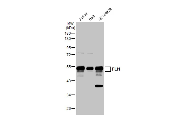
![Lane 1: Marker [kDa] 250, 130, 95, 72, 55, 36, 28, 17, 10 | Lane 2: RT4 | Lane 3: U-251 MG | Lane 4: Human Plasma | Lane 5: Liver | Lane 6: Tonsil](https://atlasantibodies.s3.amazonaws.com/images/wb/hpa065030-wb-1.jpg "Lane 1: Marker [kDa] 250, 130, 95, 72, 55, 36, 28, 17, 10 | Lane 2: RT4 | Lane 3: U-251 MG | Lane 4: Human Plasma | Lane 5: Liver | Lane 6: Tonsil")
Immunohistochemical staining of human bronchus shows cilia positivity in respiratory epithelial cells.
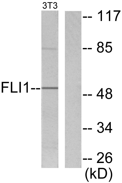
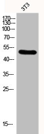
Anti-FLI1 Antibody
HPA065030
ApplicationsWestern Blot, ImmunoHistoChemistry
Product group Antibodies
ReactivityHuman
TargetFLI1
Overview
- SupplierAtlas Antibodies
- Product NameAnti-FLI1 Antibody
- Delivery Days Customer4
- ApplicationsWestern Blot, ImmunoHistoChemistry
- CertificationResearch Use Only
- ClonalityPolyclonal
- ConjugateUnconjugated
- Gene ID2313
- Target nameFLI1
- Target descriptionFli-1 proto-oncogene, ETS transcription factor
- Target synonymsBDPLT21, EWSR2, FLI-1, SIC-1, Friend leukemia integration 1 transcription factor, Ewing sarcoma breakpoint region 2, Friend leukemia virus integration 1, transcription factor ERGB
- HostRabbit
- IsotypeIgG
- Protein IDQ01543
- Protein NameFriend leukemia integration 1 transcription factor
- Scientific DescriptionRecombinant Protein Epitope Signature Tag (PrEST) antigen sequence
- ReactivityHuman
- Storage Instruction-20°C,2°C to 8°C
- UNSPSC41116161
Datasheet
MSDS
Related products
Product group Antibodies
Anti-FLI1 AntibodyA98149
ApplicationsWestern Blot, ELISA, ImmunoHistoChemistry
ReactivityHuman, Mouse
- SizePrice
Product group Antibodies
Anti-FLI1 Antibody144-05644
ApplicationsImmunoPrecipitation, Western Blot, ImmunoHistoChemistry
ReactivityHuman, Mouse, Rat
TargetFLI1
- SizePrice
Product group Antibodies
FLI1 Recombinant AntibodyBSM-60431R
ApplicationsWestern Blot
ReactivityHuman, Mouse, Rat
TargetFLI1
- SizePrice

Product group Antibodies
Anti-FLI1 Picoband(r) AntibodyA00399-CARRIER-FREE
ApplicationsFlow Cytometry, ImmunoFluorescence, Western Blot, ELISA, ImmunoCytoChemistry, ImmunoHistoChemistry
ReactivityHuman, Mouse, Rat
TargetFLI1
- SizePrice
Product group Antibodies
FLI1 AntibodyCSB-PA002539
ApplicationsWestern Blot, ELISA, ImmunoHistoChemistry
ReactivityHuman, Mouse
TargetFLI1
- SizePrice
Product group Antibodies
Fli1 Polyclonal AntibodyCAC11174
ApplicationsImmunoFluorescence, Western Blot, ELISA, ImmunoHistoChemistry
TargetFLI1
- SizePrice
Product group Antibodies

FLI1 AntibodyLS-C335594
ApplicationsImmunoFluorescence, ImmunoPrecipitation, Western Blot, ImmunoHistoChemistry
ReactivityHuman
TargetFLI1
- SizePrice

Product group Antibodies
Anti-FLI1 AntibodyHPA073099
ApplicationsChIP Chromatin ImmunoPrecipitation, ImmunoCytoChemistry
ReactivityHuman
TargetFLI1
- SizePrice
Product group Antibodies
FLI1 antibodyGTX112937
ApplicationsImmunoFluorescence, ImmunoPrecipitation, Western Blot, ImmunoCytoChemistry, ImmunoHistoChemistry, ImmunoHistoChemistry Paraffin
ReactivityHuman, Rat
TargetFLI1
- SizePrice