


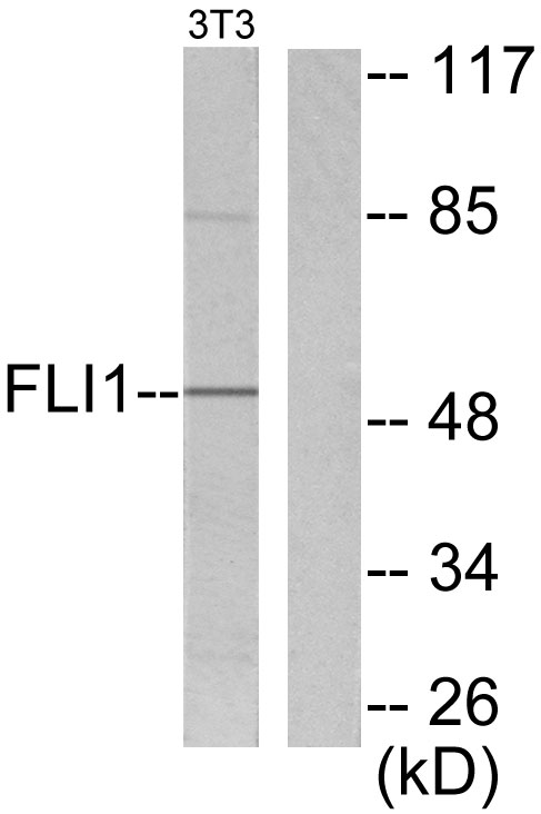
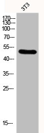
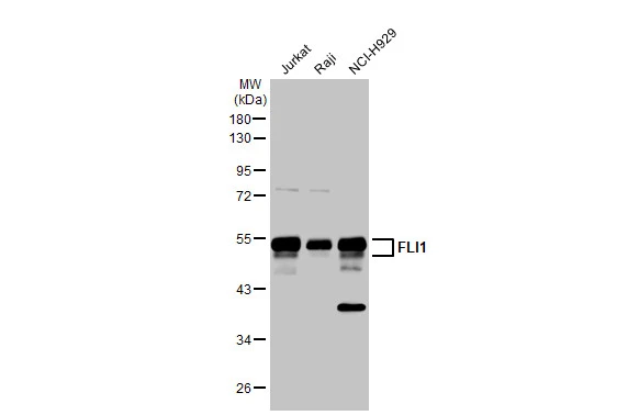
Figure 1. Western blot analysis of FLI1 using anti-FLI1 antibody (A00399). Electrophoresis was performed on a 5-20% SDS-PAGE gel at 70V (Stacking gel) / 90V (Resolving gel) for 2-3 hours. The sample well of each lane was loaded with 50ug of sample under reducing conditions. Lane 1: human Jurkat whole cell lysates, Lane 2: human HL-60 whole cell lysates, Lane 3: rat liver tissue lysates, Lane 4: mouse liver tissue lysates, Lane 5: mouse lung tissue lysates, Lane 6: mouse RAW246.7 whole cell lysates. After Electrophoresis, proteins were transferred to a Nitrocellulose membrane at 150mA for 50-90 minutes. Blocked the membrane with 5% Non-fat Milk/ TBS for 1.5 hour at RT. The membrane was incubated with rabbit anti-FLI1 antigen affinity purified polyclonal antibody (Catalog # A00399) at 0.25 microg/mL overnight at 4°C, then washed with TBS-0.1%Tween 3 times with 5 minutes each and probed with a goat anti-rabbit IgG-HRP secondary antibody at a dilution of 1:5000 for 1.5 hour at RT. The signal is developed using an Enhanced Chemiluminescent detection (ECL) kit (Catalog # EK1002) with Tanon 5200 system. A specific band was detected for FLI1 at approximately 54KD. The expected band size for FLI1 is at 51KD.
. FLI1 was detected in paraffin-embedded section of human tonsil tissue. Heat mediated antigen retrieval was performed in EDTA buffer (pH8.0, epitope retrieval solution). The tissue section was blocked with 10% goat serum. The tissue section was then incubated with 1microg/ml rabbit anti-FLI1 Antibody (A00399) overnight at 4°C. Biotinylated goat anti-rabbit IgG was used as secondary antibody and incubated for 30 minutes at 37°C. The tissue section was developed using Strepavidin-Biotin-Complex (SABC) (Catalog # SA1022) with DAB as the chromogen.")
. FLI1 was detected in immunocytochemical section of U20S cells. Enzyme antigen retrieval was performed using IHC enzyme antigen retrieval reagent (AR0022) for 15 mins. The cells were blocked with 10% goat serum. And then incubated with 2microg/mL rabbit anti-FLI1 Antibody (A00399) overnight at 4°C. Cy3 Conjugated Goat Anti-Rabbit IgG (BA1032) was used as secondary antibody at 1:100 dilution and incubated for 30 minutes at 37°C. The section was counterstained with DAPI. Visualize using a fluorescence microscope and filter sets appropriate for the label used.")
. Overlay histogram showing THP-1 cells stained with A00399 (Blue line). To facilitate intracellular staining, cells were fixed with 4% paraformaldehyde and permeabilized with permeabilization buffer. The cells were blocked with 10% normal goat serum. And then incubated with rabbit anti-FLI1 Antibody (A00399, 1microg/1x106 cells) for 30 min at 20°C. DyLight®488 conjugated goat anti-rabbit IgG (BA1127, 5-10microg/1x106 cells) was used as secondary antibody for 30 minutes at 20°C. Isotype control antibody (Green line) was rabbit IgG (1microg/1x106) used under the same conditions. Unlabelled sample without incubation with primary antibody and secondary antibody (Red line) was used as a blank control.")
Figure 1. Western blot analysis of FLI1 using anti-FLI1 antibody (A00399). Electrophoresis was performed on a 5-20% SDS-PAGE gel at 70V (Stacking gel) / 90V (Resolving gel) for 2-3 hours. The sample well of each lane was loaded with 50ug of sample under reducing conditions. Lane 1: human Jurkat whole cell lysates, Lane 2: human HL-60 whole cell lysates, Lane 3: rat liver tissue lysates, Lane 4: mouse liver tissue lysates, Lane 5: mouse lung tissue lysates, Lane 6: mouse RAW246.7 whole cell lysates. After Electrophoresis, proteins were transferred to a Nitrocellulose membrane at 150mA for 50-90 minutes. Blocked the membrane with 5% Non-fat Milk/ TBS for 1.5 hour at RT. The membrane was incubated with rabbit anti-FLI1 antigen affinity purified polyclonal antibody (Catalog # A00399) at 0.25 microg/mL overnight at 4°C, then washed with TBS-0.1%Tween 3 times with 5 minutes each and probed with a goat anti-rabbit IgG-HRP secondary antibody at a dilution of 1:5000 for 1.5 hour at RT. The signal is developed using an Enhanced Chemiluminescent detection (ECL) kit (Catalog # EK1002) with Tanon 5200 system. A specific band was detected for FLI1 at approximately 54KD. The expected band size for FLI1 is at 51KD.
Anti-FLI1 Picoband(r) Antibody
A00399-CARRIER-FREE
ApplicationsFlow Cytometry, ImmunoFluorescence, Western Blot, ELISA, ImmunoCytoChemistry, ImmunoHistoChemistry
Product group Antibodies
ReactivityHuman, Mouse, Rat
TargetFLI1
Overview
- SupplierBoster Bio
- Product NameAnti-FLI1 Picoband(r) Antibody
- Delivery Days Customer9
- Application Supplier NoteTested Species: In-house tested species with positive results. Other applications have not been tested. Optimal dilutions should be determined by end users.
- ApplicationsFlow Cytometry, ImmunoFluorescence, Western Blot, ELISA, ImmunoCytoChemistry, ImmunoHistoChemistry
- CertificationResearch Use Only
- ClonalityPolyclonal
- Concentration500 ug/ml
- Gene ID2313
- Target nameFLI1
- Target descriptionFli-1 proto-oncogene, ETS transcription factor
- Target synonymsBDPLT21, EWSR2, FLI-1, SIC-1, Friend leukemia integration 1 transcription factor, Ewing sarcoma breakpoint region 2, Friend leukemia virus integration 1, transcription factor ERGB
- HostRabbit
- IsotypeIgG
- Protein IDQ01543
- Protein NameFriend leukemia integration 1 transcription factor
- Scientific DescriptionBoster Bio Anti-FLI1 Picoband® Antibody catalog # A00399. Tested in ELISA, Flow Cytometry, IF, IHC, ICC, WB applications. This antibody reacts with Human, Mouse, Rat. The brand Picoband indicates this is a premium antibody that guarantees superior quality, high affinity, and strong signals with minimal background in Western blot applications. Only our best-performing antibodies are designated as Picoband, ensuring unmatched performance.
- ReactivityHuman, Mouse, Rat
- Storage Instruction-20°C,2°C to 8°C
- UNSPSC12352203
Related products
Product group Antibodies
Anti-FLI1 AntibodyA98149
ApplicationsWestern Blot, ELISA, ImmunoHistoChemistry
ReactivityHuman, Mouse
- SizePrice
Product group Antibodies
Anti-FLI1 Antibody144-05644
ApplicationsImmunoPrecipitation, Western Blot, ImmunoHistoChemistry
ReactivityHuman, Mouse, Rat
TargetFLI1
- SizePrice
Product group Antibodies
FLI1 Recombinant AntibodyBSM-60431R
ApplicationsWestern Blot
ReactivityHuman, Mouse, Rat
TargetFLI1
- SizePrice
Product group Antibodies
FLI1 AntibodyCSB-PA002539
ApplicationsWestern Blot, ELISA, ImmunoHistoChemistry
ReactivityHuman, Mouse
TargetFLI1
- SizePrice
Product group Antibodies
Fli1 Polyclonal AntibodyCAC11174
ApplicationsImmunoFluorescence, Western Blot, ELISA, ImmunoHistoChemistry
TargetFLI1
- SizePrice
Product group Antibodies

FLI1 AntibodyLS-C335594
ApplicationsImmunoFluorescence, ImmunoPrecipitation, Western Blot, ImmunoHistoChemistry
ReactivityHuman
TargetFLI1
- SizePrice

Product group Antibodies
Anti-FLI1 AntibodyHPA065030
ApplicationsWestern Blot, ImmunoHistoChemistry
ReactivityHuman
TargetFLI1
- SizePrice
Product group Antibodies
FLI1 antibodyGTX112937
ApplicationsImmunoFluorescence, ImmunoPrecipitation, Western Blot, ImmunoCytoChemistry, ImmunoHistoChemistry, ImmunoHistoChemistry Paraffin
ReactivityHuman, Rat
TargetFLI1
- SizePrice
Product group Antibodies
Anti-FLI1 AntibodyCAB5644
ApplicationsImmunoPrecipitation, Western Blot, ELISA
ReactivityHuman
TargetFLI1
- SizePrice