


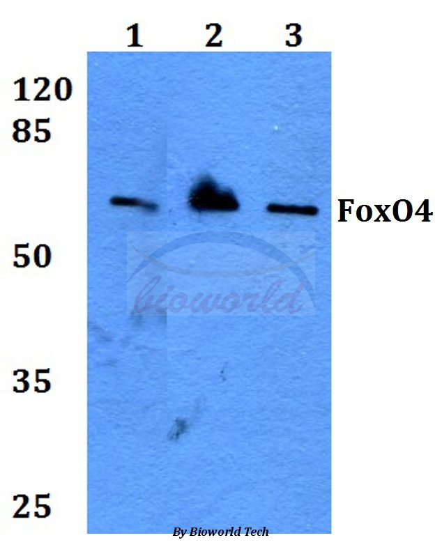
Figure 1. Western blot analysis of FOXO4 using anti-FOXO4 antibody (M01819-1). Electrophoresis was performed on a 5-20% SDS-PAGE gel at 70V (Stacking gel) / 90V (Resolving gel) for 2-3 hours. The sample well of each lane was loaded with 30 ug of sample under reducing conditions. Lane 1: human K562 whole cell lysates, Lane 2: human Hela whole cell lysates, Lane 3: human 293T whole cell lysates, Lane 4: human Caco-2 whole cell lysates, Lane 5: rat skeletal muscle tissue lysates, Lane 6: mouse heart tissue lysates. After electrophoresis, proteins were transferred to a nitrocellulose membrane at 150 mA for 50-90 minutes. Blocked the membrane with 5% non-fat milk/TBS for 1.5 hour at RT. The membrane was incubated with rabbit anti-FOXO4 antigen affinity purified monoclonal antibody (M01819-1) at 1:500 overnight at 4°C, then washed with TBS-0.1%Tween 3 times with 5 minutes each and probed with a goat anti-rabbit IgG-HRP secondary antibody at a dilution of 1:500 for 1.5 hour at RT. The signal is developed using an Enhanced Chemiluminescent detection (ECL) kit (Catalog # EK1002) with Tanon 5200 system. A specific band was detected for FOXO4 at approximately 65 kDa. The expected band size for FOXO4 is at 54 kDa.

Figure 1. Western blot analysis of FOXO4 using anti-FOXO4 antibody (M01819-1). Electrophoresis was performed on a 5-20% SDS-PAGE gel at 70V (Stacking gel) / 90V (Resolving gel) for 2-3 hours. The sample well of each lane was loaded with 30 ug of sample under reducing conditions. Lane 1: human K562 whole cell lysates, Lane 2: human Hela whole cell lysates, Lane 3: human 293T whole cell lysates, Lane 4: human Caco-2 whole cell lysates, Lane 5: rat skeletal muscle tissue lysates, Lane 6: mouse heart tissue lysates. After electrophoresis, proteins were transferred to a nitrocellulose membrane at 150 mA for 50-90 minutes. Blocked the membrane with 5% non-fat milk/TBS for 1.5 hour at RT. The membrane was incubated with rabbit anti-FOXO4 antigen affinity purified monoclonal antibody (M01819-1) at 1:500 overnight at 4°C, then washed with TBS-0.1%Tween 3 times with 5 minutes each and probed with a goat anti-rabbit IgG-HRP secondary antibody at a dilution of 1:500 for 1.5 hour at RT. The signal is developed using an Enhanced Chemiluminescent detection (ECL) kit (Catalog # EK1002) with Tanon 5200 system. A specific band was detected for FOXO4 at approximately 65 kDa. The expected band size for FOXO4 is at 54 kDa.
Anti-FoxO4/Afx Rabbit Monoclonal Antibody
M01819-1
ApplicationsFlow Cytometry, ImmunoFluorescence, ImmunoPrecipitation, Western Blot, ImmunoCytoChemistry
Product group Antibodies
ReactivityHuman, Mouse, Rat
TargetFOXO4
Overview
- SupplierBoster Bio
- Product NameAnti-FoxO4/Afx Rabbit Monoclonal Antibody
- Delivery Days Customer9
- ApplicationsFlow Cytometry, ImmunoFluorescence, ImmunoPrecipitation, Western Blot, ImmunoCytoChemistry
- CertificationResearch Use Only
- ClonalityMonoclonal
- Clone IDHEH-6
- Gene ID4303
- Target nameFOXO4
- Target descriptionforkhead box O4
- Target synonymsAFX, AFX1, MLLT7, forkhead box protein O4, fork head domain transcription factor AFX1, myeloid/lymphoid or mixed-lineage leukemia (trithorax homolog, Drosophila); translocated to, 7
- HostRabbit
- IsotypeIgG
- Protein IDP98177
- Protein NameForkhead box protein O4
- Scientific DescriptionBoster Bio Anti-FoxO4/Afx Rabbit Monoclonal Antibody catalog # M01819-1. Tested in WB, ICC/IF, IP, Flow Cytometry applications. This antibody reacts with Human, Mouse, Rat.
- ReactivityHuman, Mouse, Rat
- Storage Instruction-20°C
- UNSPSC12352203
Datasheet
MSDS
Related products
Product group Antibodies
ApplicationsWestern Blot, ImmunoHistoChemistry
ReactivityHuman, Mouse, Rat
- SizePrice
Product group Antibodies
Anti-Phospho-FOXO4-S197 Antibody144-50660
ApplicationsImmunoFluorescence, Western Blot, ImmunoHistoChemistry
ReactivityHuman, Mouse, Rat
TargetFOXO4
- SizePrice
Product group Antibodies

FOXO4 / AFX1 AntibodyLS-C832685
ApplicationsELISA, ImmunoHistoChemistry
ReactivityHuman, Mouse
TargetFOXO4
- SizePrice
Product group Antibodies
ApplicationsImmunoFluorescence, Western Blot, ELISA, ImmunoCytoChemistry, ImmunoHistoChemistry, ImmunoHistoChemistry Frozen, ImmunoHistoChemistry Paraffin
ReactivityBovine, Canine, Chicken, Equine, Human, Mouse, Porcine, Rat
TargetFOXO4
- SizePrice
Product group Antibodies
FOXO4 AntibodyCSB-PA002575
ApplicationsImmunoFluorescence, Western Blot, ELISA
ReactivityHuman, Monkey, Mouse
TargetFOXO4
- SizePrice
Product group Antibodies

Goat anti-FOXO4 / MLLT7EB07734
ApplicationsWestern Blot, ELISA
ReactivityHuman
TargetFOXO4
- SizePrice
Product group Antibodies
Anti-FOXO4 AntibodyHPA039560
ApplicationsImmunoCytoChemistry
ReactivityHuman
TargetFOXO4
- SizePrice
Product group Antibodies
FOXO4 antibodyGTX12075
ApplicationsImmunoFluorescence, ImmunoPrecipitation, Western Blot, ImmunoCytoChemistry
ReactivityHuman
TargetFOXO4
- SizePrice