


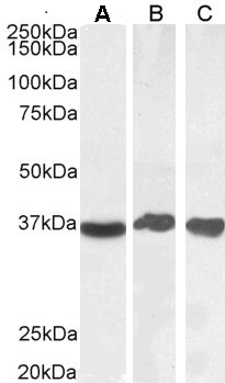
Figure 1. Western blot analysis of GAPDH using anti-GAPDH antibody (A00227-1). Electrophoresis was performed on a 5-20% SDS-PAGE gel at 70V (Stacking gel) / 90V (Resolving gel) for 2-3 hours. The sample well of each lane was loaded with 30 ug of sample under reducing conditions. Lane 1: human Hela whole cell lysates, Lane 2: human CACO-2 whole cell lysates, Lane 3: human CCRF-CEM whole cell lysates, Lane 4: rat brain tissue lysates, Lane 5: rat liver tissue lysates, Lane 6: mouse brain tissue lysates, Lane 7: mouse liver tissue lysates. After electrophoresis, proteins were transferred to a nitrocellulose membrane at 150 mA for 50-90 minutes. Blocked the membrane with 5% non-fat milk/TBS for 1.5 hour at RT. The membrane was incubated with rabbit anti-GAPDH antigen affinity purified polyclonal antibody (Catalog # A00227-1) at 0.5 microg/mL overnight at 4°C, then washed with TBS-0.1%Tween 3 times with 5 minutes each and probed with a goat anti-rabbit IgG-HRP secondary antibody at a dilution of 1:5000 for 1.5 hour at RT. The signal is developed using an Enhanced Chemiluminescent detection (ECL) kit (Catalog # EK1002) with Tanon 5200 system. A specific band was detected for GAPDH at approximately 36 kDa. The expected band size for GAPDH is at 36 kDa.
. GAPDH was detected in a paraffin-embedded section of human laryngeal squamous cell carcinoma tissue. Heat mediated antigen retrieval was performed in EDTA buffer (pH 8.0, epitope retrieval solution). The tissue section was blocked with 10% goat serum. The tissue section was then incubated with 2 microg/ml rabbit anti-GAPDH Antibody (A00227-1) overnight at 4°C. Peroxidase Conjugated Goat Anti-rabbit IgG was used as secondary antibody and incubated for 30 minutes at 37°C. The tissue section was developed using HRP Conjugated Rabbit IgG Super Vision Assay Kit (Catalog # SV0002) with DAB as the chromogen.")
. GAPDH was detected in a paraffin-embedded section of human renal clear cell carcinoma tissue. Heat mediated antigen retrieval was performed in EDTA buffer (pH 8.0, epitope retrieval solution). The tissue section was blocked with 10% goat serum. The tissue section was then incubated with 2 microg/ml rabbit anti-GAPDH Antibody (A00227-1) overnight at 4°C. Peroxidase Conjugated Goat Anti-rabbit IgG was used as secondary antibody and incubated for 30 minutes at 37°C. The tissue section was developed using HRP Conjugated Rabbit IgG Super Vision Assay Kit (Catalog # SV0002) with DAB as the chromogen.")
. GAPDH was detected in a paraffin-embedded section of human ovarian serous cancer tissue. Heat mediated antigen retrieval was performed in EDTA buffer (pH 8.0, epitope retrieval solution). The tissue section was blocked with 10% goat serum. The tissue section was then incubated with 2 microg/ml rabbit anti-GAPDH Antibody (A00227-1) overnight at 4°C. Peroxidase Conjugated Goat Anti-rabbit IgG was used as secondary antibody and incubated for 30 minutes at 37°C. The tissue section was developed using HRP Conjugated Rabbit IgG Super Vision Assay Kit (Catalog # SV0002) with DAB as the chromogen.")
. GAPDH was detected in an immunocytochemical section of A549 cells. Enzyme antigen retrieval was performed using IHC enzyme antigen retrieval reagent (AR0022) for 15 mins. The cells were blocked with 10% goat serum. And then incubated with 5 microg/mL rabbit anti-GAPDH Antibody (A00227-1) overnight at 4°C. DyLight®488 Conjugated Goat Anti-Rabbit IgG (BA1127) was used as secondary antibody at 1:500 dilution and incubated for 30 minutes at 37°C. The section was counterstained with DAPI. Visualize using a fluorescence microscope and filter sets appropriate for the label used.")
. Overlay histogram showing Hela cells stained with A00227-1 (Blue line). To facilitate intracellular staining, cells were fixed with 4% paraformaldehyde and permeabilized with permeabilization buffer. The cells were blocked with 10% normal goat serum. And then incubated with rabbit anti-GAPDH Antibody (A00227-1, 1 microg/1x106 cells) for 30 min at 20°C. DyLight®488 conjugated goat anti-rabbit IgG (BA1127, 5-10 microg/1x106 cells) was used as secondary antibody for 30 minutes at 20°C. Isotype control antibody (Green line) was rabbit IgG (1 microg/1x106) used under the same conditions. Unlabelled sample without incubation with primary antibody and secondary antibody (Red line) was used as a blank control.")
Figure 1. Western blot analysis of GAPDH using anti-GAPDH antibody (A00227-1). Electrophoresis was performed on a 5-20% SDS-PAGE gel at 70V (Stacking gel) / 90V (Resolving gel) for 2-3 hours. The sample well of each lane was loaded with 30 ug of sample under reducing conditions. Lane 1: human Hela whole cell lysates, Lane 2: human CACO-2 whole cell lysates, Lane 3: human CCRF-CEM whole cell lysates, Lane 4: rat brain tissue lysates, Lane 5: rat liver tissue lysates, Lane 6: mouse brain tissue lysates, Lane 7: mouse liver tissue lysates. After electrophoresis, proteins were transferred to a nitrocellulose membrane at 150 mA for 50-90 minutes. Blocked the membrane with 5% non-fat milk/TBS for 1.5 hour at RT. The membrane was incubated with rabbit anti-GAPDH antigen affinity purified polyclonal antibody (Catalog # A00227-1) at 0.5 microg/mL overnight at 4°C, then washed with TBS-0.1%Tween 3 times with 5 minutes each and probed with a goat anti-rabbit IgG-HRP secondary antibody at a dilution of 1:5000 for 1.5 hour at RT. The signal is developed using an Enhanced Chemiluminescent detection (ECL) kit (Catalog # EK1002) with Tanon 5200 system. A specific band was detected for GAPDH at approximately 36 kDa. The expected band size for GAPDH is at 36 kDa.
Anti-GAPDH Antibody Picoband(r)
A00227-1-CARRIER-FREE
ApplicationsFlow Cytometry, ImmunoFluorescence, Western Blot, ImmunoCytoChemistry, ImmunoHistoChemistry
Product group Antibodies
ReactivityChicken, Human, Monkey, Mouse, Rat, Zebra Fish
TargetGAPDH
Overview
- SupplierBoster Bio
- Product NameAnti-GAPDH Antibody Picoband(r)
- Delivery Days Customer9
- ApplicationsFlow Cytometry, ImmunoFluorescence, Western Blot, ImmunoCytoChemistry, ImmunoHistoChemistry
- CertificationResearch Use Only
- ClonalityPolyclonal
- Concentration500 ug/ml
- Gene ID2597
- Target nameGAPDH
- Target descriptionglyceraldehyde-3-phosphate dehydrogenase
- Target synonymsG3PD, GAPD, HEL-S-162eP, glyceraldehyde-3-phosphate dehydrogenase, OCAS, p38 component, Oct1 coactivator in S phase, 38 Kd component, aging-associated gene 9 protein, epididymis secretory sperm binding protein Li 162eP, peptidyl-cysteine S-nitrosylase GAPDH
- HostRabbit
- IsotypeIgG
- Protein IDP04406
- Protein NameGlyceraldehyde-3-phosphate dehydrogenase
- Scientific DescriptionBoster Bio Anti-GAPDH Antibody Picoband® catalog # A00227-1. Tested in Flow Cytometry, IF, IHC, ICC, WB applications. This antibody reacts with Human, Mouse, Rat, Monkey, Chicken, Zebrafish. The brand Picoband indicates this is a premium antibody that guarantees superior quality, high affinity, and strong signals with minimal background in Western blot applications. Only our best-performing antibodies are designated as Picoband, ensuring unmatched performance.
- ReactivityChicken, Human, Monkey, Mouse, Rat, Zebra Fish
- Storage Instruction-20°C,2°C to 8°C
- UNSPSC12352203
Related products
Product group Antibodies
Anti-GAPDH AntibodyA82908
ApplicationsImmunoFluorescence, Western Blot, ELISA, ImmunoHistoChemistry
ReactivityHuman, Mouse, Rat
- SizePrice
Product group Antibodies
Anti-GAPDH (63~83aa) Antibody130-10873
ApplicationsELISA
ReactivityHuman, Rabbit
TargetGAPDH
- SizePrice


Product group Antibodies
Anti-GAPDH AntibodyAMAB91152
ApplicationsWestern Blot, ImmunoHistoChemistry
ReactivityHuman, Mouse, Rat
TargetGAPDH
- SizePrice

Product group Antibodies
References
GAPDH Polyclonal AntibodyBS-10900R
ApplicationsImmunoFluorescence, Western Blot, ImmunoCytoChemistry, ImmunoHistoChemistry, ImmunoHistoChemistry Frozen, ImmunoHistoChemistry Paraffin
TargetGAPDH
- SizePrice
Product group Antibodies
GAPDH Monoclonal AntibodyCSB-MA000184
ApplicationsWestern Blot, ELISA, ImmunoHistoChemistry
ReactivityCanine, Chicken, Hamster, Human, Insect, Monkey, Mouse, Porcine, Rabbit, Rat, Sheep, Yeast
TargetGAPDH
- SizePrice
Product group Antibodies

ApplicationsWestern Blot, ELISA
ReactivityCanine, Human, Mouse, Porcine, Rat
TargetGAPDH
- SizePrice
Product group Antibodies
Gapdh Polyclonal AntibodyCAC07001
ApplicationsImmunoFluorescence, Western Blot, ELISA, ImmunoHistoChemistry
TargetGAPDH
- SizePrice
Product group Antibodies
GAPDH antibodyGTX100118
ApplicationsImmunoFluorescence, ImmunoPrecipitation, Western Blot, ImmunoCytoChemistry, ImmunoHistoChemistry, ImmunoHistoChemistry Paraffin
ReactivityBacteria, Bovine, Canine, C. Elegans, Chicken, Drosophila, Fish, Hamster, Human, Insect, Mammals, Monkey, Mouse, Plant, Porcine, Rabbit, Rat, Zebra Fish, Other Species
TargetGAPDH
- SizePrice