

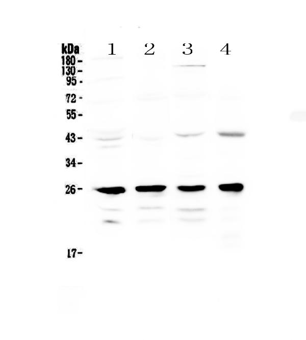
Figure 1. Western blot analysis of GSTM1 using anti-GSTM1 antibody (A00569-1). Electrophoresis was performed on a 5-20% SDS-PAGE gel at 70V (Stacking gel) / 90V (Resolving gel) for 2-3 hours. The sample well of each lane was loaded with 50ug of sample under reducing conditions. Lane 1: rat brain tissue lysates, Lane 2: rat lung tissue lysates, Lane 3: mouse stomach tissue lysates, Lane 4: mouse kidney tissue lysates. After Electrophoresis, proteins were transferred to a Nitrocellulose membrane at 150mA for 50-90 minutes. Blocked the membrane with 5% Non-fat Milk/ TBS for 1.5 hour at RT. The membrane was incubated with rabbit anti-GSTM1 antigen affinity purified polyclonal antibody (Catalog # A00569-1) at 0.5 microg/mL overnight at 4°C, then washed with TBS-0.1%Tween 3 times with 5 minutes each and probed with a goat anti-rabbit IgG-HRP secondary antibody at a dilution of 1:10000 for 1.5 hour at RT. The signal is developed using an Enhanced Chemiluminescent detection (ECL) kit (Catalog # EK1002) with Tanon 5200 system. A specific band was detected for GSTM1 at approximately 26KD. The expected band size for GSTM1 is at 26KD.
. Overlay histogram showing HeLa cells stained with A00569-1 (Blue line). To facilitate intracellular staining, cells were fixed with 4% paraformaldehyde and permeabilized with permeabilization buffer. The cells were blocked with 10% normal goat serum. And then incubated with rabbit anti-GSTM1 Antibody (A00569-1,1microg/1x106 cells) for 30 min at 20°C. DyLight®488 conjugated goat anti-rabbit IgG (BA1127, 5-10microg/1x106 cells) was used as secondary antibody for 30 minutes at 20°C. Isotype control antibody (Green line) was rabbit IgG (1microg/1x106) used under the same conditions. Unlabelled sample without incubation with primary antibody and secondary antibody (Red line) was used as a blank control.")
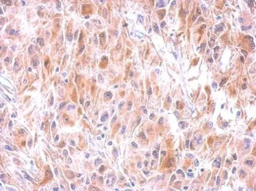
. GSTM1 was detected in an immunocytochemical section of U20S cells. Enzyme antigen retrieval was performed using IHC enzyme antigen retrieval reagent (AR0022) for 15 mins. The cells were blocked with 10% goat serum. And then incubated with 5 microg/mL rabbit anti-GSTM1 Antibody (A00569-1) overnight at 4°C. DyLight®488 Conjugated Goat Anti-Rabbit IgG (BA1127) was used as secondary antibody at 1:500 dilution and incubated for 30 minutes at 37°C. The section was counterstained with DAPI. Visualize using a fluorescence microscope and filter sets appropriate for the label used.")
Figure 1. Western blot analysis of GSTM1 using anti-GSTM1 antibody (A00569-1). Electrophoresis was performed on a 5-20% SDS-PAGE gel at 70V (Stacking gel) / 90V (Resolving gel) for 2-3 hours. The sample well of each lane was loaded with 50ug of sample under reducing conditions. Lane 1: rat brain tissue lysates, Lane 2: rat lung tissue lysates, Lane 3: mouse stomach tissue lysates, Lane 4: mouse kidney tissue lysates. After Electrophoresis, proteins were transferred to a Nitrocellulose membrane at 150mA for 50-90 minutes. Blocked the membrane with 5% Non-fat Milk/ TBS for 1.5 hour at RT. The membrane was incubated with rabbit anti-GSTM1 antigen affinity purified polyclonal antibody (Catalog # A00569-1) at 0.5 microg/mL overnight at 4°C, then washed with TBS-0.1%Tween 3 times with 5 minutes each and probed with a goat anti-rabbit IgG-HRP secondary antibody at a dilution of 1:10000 for 1.5 hour at RT. The signal is developed using an Enhanced Chemiluminescent detection (ECL) kit (Catalog # EK1002) with Tanon 5200 system. A specific band was detected for GSTM1 at approximately 26KD. The expected band size for GSTM1 is at 26KD.
Anti-GSTM1 Antibody Picoband(r)
A00569-1-CARRIER-FREE
ApplicationsFlow Cytometry, ImmunoFluorescence, Western Blot, ImmunoCytoChemistry
Product group Antibodies
ReactivityHuman, Mouse, Rat
TargetGSTM1
Overview
- SupplierBoster Bio
- Product NameAnti-GSTM1 Antibody Picoband(r)
- Delivery Days Customer9
- ApplicationsFlow Cytometry, ImmunoFluorescence, Western Blot, ImmunoCytoChemistry
- CertificationResearch Use Only
- ClonalityPolyclonal
- Concentration500 ug/ml
- Gene ID2944
- Target nameGSTM1
- Target descriptionglutathione S-transferase mu 1
- Target synonymsGST1, GSTM1-1, GSTM1a-1a, GSTM1b-1b, GTH4, GTM1, H-B, MU, MU-1, glutathione S-transferase Mu 1, GST HB subunit 4, GST class-mu 1, HB subunit 4, S-(hydroxyalkyl)glutathione lyase, glutathione S-alkyltransferase, glutathione S-aralkyltransferase, glutathione S-aryltransferase, glutathione S-transferase M1
- HostRabbit
- IsotypeIgG
- Protein IDP09488
- Protein NameGlutathione S-transferase Mu 1
- Scientific DescriptionBoster Bio Anti-GSTM1 Antibody Picoband® catalog # A00569-1. Tested in Flow Cytometry, IF, ICC, WB applications. This antibody reacts with Human, Mouse, Rat. The brand Picoband indicates this is a premium antibody that guarantees superior quality, high affinity, and strong signals with minimal background in Western blot applications. Only our best-performing antibodies are designated as Picoband, ensuring unmatched performance.
- ReactivityHuman, Mouse, Rat
- Storage Instruction-20°C,2°C to 8°C
- UNSPSC12352203
Related products
Product group Antibodies
Anti-GSTM1 AntibodyA97496
ApplicationsWestern Blot, ELISA
ReactivityHuman, Mouse, Rat
- SizePrice
Product group Antibodies
Anti-GSTM1 Antibody144-64951
ApplicationsImmunoFluorescence, Western Blot
ReactivityHuman, Mouse, Rat
TargetGSTM1
- SizePrice
Product group Antibodies

GSTM1 AntibodyLS-C831579
ApplicationsWestern Blot
ReactivityRat
TargetGSTM1
- SizePrice
Product group Antibodies
GST1 Polyclonal AntibodyBS-0663R
ApplicationsImmunoFluorescence, Western Blot, ImmunoCytoChemistry, ImmunoHistoChemistry, ImmunoHistoChemistry Frozen, ImmunoHistoChemistry Paraffin
ReactivityHuman
TargetGSTM1
- SizePrice
Product group Antibodies
GSTM1 AntibodyCSB-PA009979LA01HU
ApplicationsImmunoFluorescence, ELISA, ImmunoHistoChemistry
ReactivityHuman
TargetGSTM1
- SizePrice
Product group Antibodies
References

Goat anti-GSTM1 / GSTM2EB06606
ApplicationsWestern Blot, ELISA
ReactivityHuman
TargetGSTM1
- SizePrice
Product group Antibodies
ApplicationsImmunoPrecipitation, Western Blot, ImmunoCytoChemistry, ImmunoHistoChemistry
ReactivityMouse, Porcine, Rat
TargetGSTM1
- SizePrice
Product group Antibodies
GSTM1 antibodyGTX100298
ApplicationsWestern Blot, ImmunoHistoChemistry, ImmunoHistoChemistry Paraffin
ReactivityHuman
TargetGSTM1
- SizePrice

Product group Antibodies
Anti-GSTM1 AntibodyHPA048652
ApplicationsImmunoHistoChemistry
ReactivityHuman
TargetGSTM1
- SizePrice
Product group Antibodies
TargetGSTM1
- SizePrice