


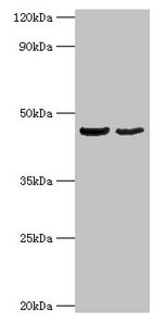
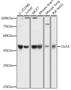
Figure 1. Western blot analysis of GTPBP9/OLA1 using anti-GTPBP9/OLA1 antibody (A07162). Electrophoresis was performed on a 5-20% SDS-PAGE gel at 70V (Stacking gel) / 90V (Resolving gel) for 2-3 hours. The sample well of each lane was loaded with 30 ug of sample under reducing conditions. Lane 1: human HepG2 whole cell lysates, Lane 2: human Jurkat whole cell lysates, Lane 3: human MCF-7 whole cell lysates, Lane 4: human PC-3 whole cell lysates, Lane 5: rat brain tissue lysates, Lane 6: rat liver tissue lysates, Lane 7: mouse brain tissue lysates, Lane 8: mouse liver tissue lysates. After electrophoresis, proteins were transferred to a nitrocellulose membrane at 150 mA for 50-90 minutes. Blocked the membrane with 5% non-fat milk/TBS for 1.5 hour at RT. The membrane was incubated with rabbit anti-GTPBP9/OLA1 antigen affinity purified polyclonal antibody (Catalog # A07162) at 0.25 microg/mL overnight at 4°C, then washed with TBS-0.1%Tween 3 times with 5 minutes each and probed with a goat anti-rabbit IgG-HRP secondary antibody at a dilution of 1:5000 for 1.5 hour at RT. The signal is developed using an Enhanced Chemiluminescent detection (ECL) kit (Catalog # EK1002) with Tanon 5200 system. A specific band was detected for GTPBP9/OLA1 at approximately 45 kDa. The expected band size for GTPBP9/OLA1 is at 45 kDa.
. GTPBP9/OLA1 was detected in an immunocytochemical section of U2OS cells. Enzyme antigen retrieval was performed using IHC enzyme antigen retrieval reagent (AR0022) for 15 mins. The cells were blocked with 10% goat serum. And then incubated with 5 microg/mL rabbit anti-GTPBP9/OLA1 Antibody (A07162) overnight at 4°C. Cy3 Conjugated Goat Anti-Rabbit IgG (BA1032) was used as secondary antibody at 1:500 dilution and incubated for 30 minutes at 37°C. The section was counterstained with DAPI. Visualize using a fluorescence microscope and filter sets appropriate for the label used.")
. Overlay histogram showing Raji cells stained with A07162 (Blue line). To facilitate intracellular staining, cells were fixed with 4% paraformaldehyde and permeabilized with permeabilization buffer. The cells were blocked with 10% normal goat serum. And then incubated with rabbit anti-GTPBP9/OLA1 Antibody (A07162, 1 microg/1x106 cells) for 30 min at 20°C. DyLight®488 conjugated goat anti-rabbit IgG (BA1127, 5-10 microg/1x106 cells) was used as secondary antibody for 30 minutes at 20°C. Isotype control antibody (Green line) was rabbit IgG (1 microg/1x106) used under the same conditions. Unlabelled sample without incubation with primary antibody and secondary antibody (Red line) was used as a blank control.")
Figure 1. Western blot analysis of GTPBP9/OLA1 using anti-GTPBP9/OLA1 antibody (A07162). Electrophoresis was performed on a 5-20% SDS-PAGE gel at 70V (Stacking gel) / 90V (Resolving gel) for 2-3 hours. The sample well of each lane was loaded with 30 ug of sample under reducing conditions. Lane 1: human HepG2 whole cell lysates, Lane 2: human Jurkat whole cell lysates, Lane 3: human MCF-7 whole cell lysates, Lane 4: human PC-3 whole cell lysates, Lane 5: rat brain tissue lysates, Lane 6: rat liver tissue lysates, Lane 7: mouse brain tissue lysates, Lane 8: mouse liver tissue lysates. After electrophoresis, proteins were transferred to a nitrocellulose membrane at 150 mA for 50-90 minutes. Blocked the membrane with 5% non-fat milk/TBS for 1.5 hour at RT. The membrane was incubated with rabbit anti-GTPBP9/OLA1 antigen affinity purified polyclonal antibody (Catalog # A07162) at 0.25 microg/mL overnight at 4°C, then washed with TBS-0.1%Tween 3 times with 5 minutes each and probed with a goat anti-rabbit IgG-HRP secondary antibody at a dilution of 1:5000 for 1.5 hour at RT. The signal is developed using an Enhanced Chemiluminescent detection (ECL) kit (Catalog # EK1002) with Tanon 5200 system. A specific band was detected for GTPBP9/OLA1 at approximately 45 kDa. The expected band size for GTPBP9/OLA1 is at 45 kDa.
Anti-GTPBP9/OLA1 Antibody Picoband(r)
A07162-CARRIER-FREE
ApplicationsFlow Cytometry, ImmunoFluorescence, Western Blot, ELISA, ImmunoCytoChemistry
Product group Antibodies
ReactivityHuman, Mouse, Rat
TargetOLA1
Overview
- SupplierBoster Bio
- Product NameAnti-GTPBP9/OLA1 Antibody Picoband(r)
- Delivery Days Customer9
- Application Supplier NoteTested Species: In-house tested species with positive results. Other applications have not been tested. Optimal dilutions should be determined by end users.
- ApplicationsFlow Cytometry, ImmunoFluorescence, Western Blot, ELISA, ImmunoCytoChemistry
- CertificationResearch Use Only
- ClonalityPolyclonal
- Concentration500 ug/ml
- Gene ID29789
- Target nameOLA1
- Target descriptionObg like ATPase 1
- Target synonymsDOC45, GBP45, GTBP9, GTPBP9, PTD004, obg-like ATPase 1, DNA damage-regulated overexpressed in cancer 45 protein, GTP-binding protein 9 (putative), GTP-binding protein PTD004, homologous yeast-44.2 protein
- HostRabbit
- IsotypeIgG
- Protein IDQ9NTK5
- Protein NameObg-like ATPase 1
- Scientific DescriptionBoster Bio Anti-GTPBP9/OLA1 Antibody Picoband® catalog # A07162. Tested in ELISA, Flow Cytometry, IF, ICC, WB applications. This antibody reacts with Human, Mouse, Rat. The brand Picoband indicates this is a premium antibody that guarantees superior quality, high affinity, and strong signals with minimal background in Western blot applications. Only our best-performing antibodies are designated as Picoband, ensuring unmatched performance.
- ReactivityHuman, Mouse, Rat
- Storage Instruction-20°C,2°C to 8°C
- UNSPSC12352203
References
- Koller-Eichhorn R, Marquardt T, Gail R, et al. Human OLA1 defines an ATPase subfamily in the Obg family of GTP-binding proteins. J Biol Chem. 2007,282(27):19928-37.Read this paper
Related products
Product group Antibodies
Anti-OLA1 Antibody144-62102
ApplicationsWestern Blot
ReactivityHuman, Mouse, Rat
TargetOLA1
- SizePrice
Product group Antibodies
OLA1 Polyclonal AntibodyCAC13852
ApplicationsImmunoFluorescence, Western Blot, ELISA, ImmunoHistoChemistry
ReactivityMouse
TargetOLA1
- SizePrice
Product group Antibodies
GTPBP9 Polyclonal AntibodyBS-9554R
ApplicationsImmunoFluorescence, Western Blot, ImmunoHistoChemistry, ImmunoHistoChemistry Paraffin
ReactivityBovine, Canine, Drosophila, Human, Mouse, Rat, Sheep
TargetOLA1
- SizePrice
Product group Antibodies
OLA1 AntibodyCSB-PA016318HA01HU
ApplicationsImmunoFluorescence, Western Blot, ELISA, ImmunoHistoChemistry
ReactivityHuman, Mouse
TargetOLA1
- SizePrice
Product group Antibodies
Anti-OLA1 AntibodyA81079
ApplicationsImmunoFluorescence, Western Blot, ImmunoCytoChemistry, ImmunoHistoChemistry
ReactivityHuman, Mouse, Rat
- SizePrice
Product group Antibodies

OLA1 AntibodyLS-C830511
ApplicationsELISA, ImmunoHistoChemistry
ReactivityHuman, Mouse, Rat
TargetOLA1
- SizePrice

Product group Antibodies
Anti-OLA1 AntibodyHPA035790
ApplicationsWestern Blot, ImmunoCytoChemistry, ImmunoHistoChemistry
ReactivityHuman, Mouse, Rat
TargetOLA1
- SizePrice
Product group Antibodies
GTPBP9 antibody [N1C1]GTX118604
ApplicationsImmunoFluorescence, Western Blot, ImmunoCytoChemistry, ImmunoHistoChemistry, ImmunoHistoChemistry Paraffin
ReactivityHuman, Mouse, Rat
TargetOLA1
- SizePrice