


Anti-OLA1 Antibody
A81079
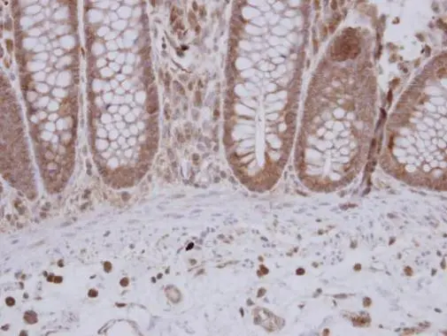
ApplicationsImmunoFluorescence, Western Blot, ImmunoCytoChemistry, ImmunoHistoChemistry
Product group Antibodies
ReactivityHuman, Mouse, Rat
Overview
- SupplierAntibodies.com
- Product NameAnti-OLA1 Antibody
- Delivery Days Customer7
- ApplicationsImmunoFluorescence, Western Blot, ImmunoCytoChemistry, ImmunoHistoChemistry
- CertificationResearch Use Only
- ClonalityPolyclonal
- ConjugateUnconjugated
- HostRabbit
- IsotypeIgG
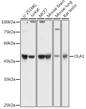
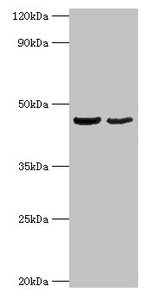
- Scientific DescriptionRabbit polyclonal antibody to OLA1.
- ReactivityHuman, Mouse, Rat
- UNSPSC12352203
Related products
Product group Antibodies
Anti-OLA1 Antibody144-62102
ApplicationsWestern Blot
ReactivityHuman, Mouse, Rat
TargetOLA1
- SizePrice
Product group Antibodies
OLA1 Polyclonal AntibodyCAC13852
ApplicationsImmunoFluorescence, Western Blot, ELISA, ImmunoHistoChemistry
ReactivityMouse
TargetOLA1
- SizePrice
Product group Antibodies
GTPBP9 Polyclonal AntibodyBS-9554R
ApplicationsImmunoFluorescence, Western Blot, ImmunoHistoChemistry, ImmunoHistoChemistry Paraffin
ReactivityBovine, Canine, Drosophila, Human, Mouse, Rat, Sheep
TargetOLA1
- SizePrice
Product group Antibodies
OLA1 AntibodyCSB-PA016318HA01HU
ApplicationsImmunoFluorescence, Western Blot, ELISA, ImmunoHistoChemistry
ReactivityHuman, Mouse
TargetOLA1
- SizePrice
Product group Antibodies

OLA1 AntibodyLS-C830511
ApplicationsELISA, ImmunoHistoChemistry
ReactivityHuman, Mouse, Rat
TargetOLA1
- SizePrice

Product group Antibodies
Anti-OLA1 AntibodyHPA035790
ApplicationsWestern Blot, ImmunoCytoChemistry, ImmunoHistoChemistry
ReactivityHuman, Mouse, Rat
TargetOLA1
- SizePrice
Product group Antibodies
GTPBP9 antibody [N1C1]GTX118604
ApplicationsImmunoFluorescence, Western Blot, ImmunoCytoChemistry, ImmunoHistoChemistry, ImmunoHistoChemistry Paraffin
ReactivityHuman, Mouse, Rat
TargetOLA1
- SizePrice

Product group Antibodies
References
Anti-GTPBP9/OLA1 Antibody Picoband(r)A07162-CARRIER-FREE
ApplicationsFlow Cytometry, ImmunoFluorescence, Western Blot, ELISA, ImmunoCytoChemistry
ReactivityHuman, Mouse, Rat
TargetOLA1
- SizePrice