

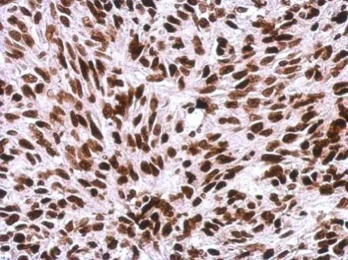
Immunohistochemical analysis of paraffin-embedded SkHep1 xenograft, using Histone H1.0(GTX114462) antibody at 1:500 dilution.
Antigen Retrieval: Trilogy? (EDTA based, pH 8.0) buffer, 15min
 was separated by 12% SDS-PAGE, and the membrane was blotted with Histone H1 antibody (GTX114462) diluted by 1:5000. The HRP-conjugated anti-rabbit IgG antibody (GTX213110-01) was used to detect the primary antibody.")
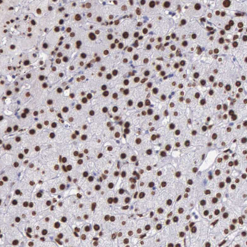
 dilution: 1:500.
Antigen Retrieval: Trilogy? (EDTA based, pH 8.0) buffer, 15min")
 were separated by 12% SDS-PAGE, and the membrane was blotted with Histone H1 antibody (GTX114462) at a dilution of 1:10000 and developed with Trident femto Western HRP Substrate (GTX14698). The HRP-conjugated anti-rabbit IgG antibody (GTX213110-01) was used to detect the primary antibody.")
 diluted at 1:500.
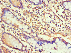
Antigen Retrieval: Citrate buffer, pH 6.0, 15 min")
 antibody at 1:500 dilution.
Antigen Retrieval: Trilogy? (EDTA based, pH 8.0) buffer, 15min")
![Histone H1.0 antibody detects Histone H1.0 protein at nucleus by immunofluorescent analysis. Sample: 293T cells were fixed in 4% paraformaldehyde at RT for 15 min. Green: Histone H1.0 stained by Histone H1.0 antibody (GTX114462) diluted at 1:500. Red: alpha Tubulin, a cytoskeleton marker, stained by alpha Tubulin antibody [GT114] (GTX628802) diluted at 1:1000.](https://www.genetex.com/upload/website/prouct_img/normal/GTX114462/GTX114462_44223_20220121_ICC_IF_w_23060501_767.webp "Histone H1.0 antibody detects Histone H1.0 protein at nucleus by immunofluorescent analysis. Sample: 293T cells were fixed in 4% paraformaldehyde at RT for 15 min. Green: Histone H1.0 stained by Histone H1.0 antibody (GTX114462) diluted at 1:500. Red: alpha Tubulin, a cytoskeleton marker, stained by alpha Tubulin antibody [GT114] (GTX628802) diluted at 1:1000.")
 diluted at 1:500.
Antigen Retrieval: Citrate buffer, pH 6.0, 15 min")
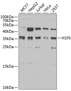
 were separated by 12% SDS-PAGE, and the membrane was blotted with Histone H1.0 antibody (GTX114462) diluted at 1:10000. The HRP-conjugated anti-rabbit IgG antibody (GTX213110-01) was used to detect the primary antibody.")
 diluted at 1:1000. Red: phalloidin, a cytoskeleton marker, diluted at 1:200. Scale bar= 10 μm.")
Immunohistochemical analysis of paraffin-embedded SkHep1 xenograft, using Histone H1.0(GTX114462) antibody at 1:500 dilution.
Antigen Retrieval: Trilogy? (EDTA based, pH 8.0) buffer, 15min
Histone H1.0 antibody
GTX114462
ApplicationsImmunoFluorescence, Western Blot, ImmunoCytoChemistry, ImmunoHistoChemistry, ImmunoHistoChemistry Paraffin
Product group Antibodies
ReactivityHuman, Mouse, Rat
TargetH1-0
Overview
- SupplierGeneTex
- Product NameHistone H1.0 antibody
- Delivery Days Customer9
- Application Supplier NoteWB: 1:1000-1:20000. ICC/IF: 1:100-1:1000. IHC-P: 1:100-1:1000. *Optimal dilutions/concentrations should be determined by the researcher.Not tested in other applications.
- ApplicationsImmunoFluorescence, Western Blot, ImmunoCytoChemistry, ImmunoHistoChemistry, ImmunoHistoChemistry Paraffin
- CertificationResearch Use Only
- ClonalityPolyclonal
- Concentration0.53 mg/ml
- ConjugateUnconjugated
- Gene ID3005
- Target nameH1-0
- Target descriptionH1.0 linker histone
- Target synonymsH1.0, H10, H1F0, H1FV, histone H1.0, H1 histone family member 0, H1.0, H1(0), H1-0, histone H1', histone H1(0)
- HostRabbit
- IsotypeIgG
- Protein IDP07305
- Protein NameHistone H1.0
- Scientific DescriptionHistones are basic nuclear proteins that are responsible for the nucleosome structure of the chromosomal fiber in eukaryotes. Nucleosomes consist of approximately 146 bp of DNA wrapped around a histone octamer composed of pairs of each of the four core histones (H2A, H2B, H3, and H4). The chromatin fiber is further compacted through the interaction of a linker histone, H1, with the DNA between the nucleosomes to form higher order chromatin structures. This gene is intronless and encodes a member of the histone H1 family. [provided by RefSeq]
- ReactivityHuman, Mouse, Rat
- Storage Instruction-20°C or -80°C,2°C to 8°C
- UNSPSC41116161
Datasheet
Related products
Product group Antibodies
ApplicationsImmunoFluorescence, Western Blot, ImmunoCytoChemistry, ImmunoHistoChemistry
ReactivityHuman, Mouse, Rat
- SizePrice


Product group Antibodies
Anti-Histone H1.0/H1F0 Antibody Picoband(r)A08821-1-CARRIER-FREE
ApplicationsFlow Cytometry, ImmunoFluorescence, Western Blot, ELISA, ImmunoCytoChemistry, ImmunoHistoChemistry
ReactivityHuman, Mouse, Rat
TargetH1-0
- SizePrice
Product group Antibodies
Anti-H1F0 Antibody144-03298
ApplicationsImmunoFluorescence, Western Blot, ImmunoHistoChemistry
ReactivityHuman, Mouse, Rat
TargetH1-0
- SizePrice
Product group Antibodies
Histone H1.0 Polyclonal AntibodyBS-33535R
ApplicationsWestern Blot, ELISA
ReactivityHuman, Mouse, Rat
TargetH1-0
- SizePrice
Product group Antibodies
H1F0 AntibodyCSB-PA010087ESR1HU
ApplicationsELISA, ImmunoHistoChemistry
ReactivityHuman
TargetH1-0
- SizePrice
Product group Antibodies
H1F0 (Ab-11) Polyclonal AntibodyCAC15243
ApplicationsWestern Blot, ELISA
ReactivityRat
TargetH1-0
- SizePrice
Product group Antibodies

H1F0 AntibodyLS-C409090
ApplicationsWestern Blot
ReactivityHuman
TargetH1-0
- SizePrice
Product group Antibodies
Anti-H1F0 AntibodyHPA000843
ApplicationsWestern Blot, ImmunoCytoChemistry, ImmunoHistoChemistry
ReactivityHuman
TargetH1-0
- SizePrice