


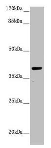
Figure 1. Western blot analysis of H2AFY/MACROH2A1 using anti-H2AFY/MACROH2A1 antibody (A04635-3). Electrophoresis was performed on a 5-20% SDS-PAGE gel at 70V (Stacking gel) / 90V (Resolving gel) for 2-3 hours. The sample well of each lane was loaded with 30 ug of sample under reducing conditions. Lane 1: human A549 whole cell lysates, Lane 2: human Caco-2 whole cell lysates, Lane 3: human Hela whole cell lysates, Lane 4: human 293T whole cell lysates, Lane 5: rat brain tissue lysates, Lane 6: rat thymus tissue lysates, Lane 7: mouse brain tissue lysates, Lane 8: mouse thymus tissue lysates. After electrophoresis, proteins were transferred to a nitrocellulose membrane at 150 mA for 50-90 minutes. Blocked the membrane with 5% non-fat milk/TBS for 1.5 hour at RT. The membrane was incubated with rabbit anti-H2AFY/MACROH2A1 antigen affinity purified polyclonal antibody (Catalog # A04635-3) at 0.5 microg/mL overnight at 4°C, then washed with TBS-0.1%Tween 3 times with 5 minutes each and probed with a goat anti-rabbit IgG-HRP secondary antibody at a dilution of 1:5000 for 1.5 hour at RT. The signal is developed using an Enhanced Chemiluminescent detection (ECL) kit (Catalog # EK1002) with Tanon 5200 system. A specific band was detected for H2AFY/MACROH2A1 at approximately 39 kDa. The expected band size for H2AFY/MACROH2A1 is at 40 kDa.
. H2AFY/MACROH2A1 was detected in a paraffin-embedded section of human colon adenocarcinoma tissue. Heat mediated antigen retrieval was performed in EDTA buffer (pH 8.0, epitope retrieval solution). The tissue section was blocked with 10% goat serum. The tissue section was then incubated with 2 microg/ml rabbit anti-H2AFY/MACROH2A1 Antibody (A04635-3) overnight at 4°C. Peroxidase Conjugated Goat Anti-rabbit IgG was used as secondary antibody and incubated for 30 minutes at 37°C. The tissue section was developed using HRP Conjugated Rabbit IgG Super Vision Assay Kit (Catalog # SV0002) with DAB as the chromogen.")
. H2AFY/MACROH2A1 was detected in a paraffin-embedded section of human colon adenocarcinoma tissue. Heat mediated antigen retrieval was performed in EDTA buffer (pH 8.0, epitope retrieval solution). The tissue section was blocked with 10% goat serum. The tissue section was then incubated with 2 microg/ml rabbit anti-H2AFY/MACROH2A1 Antibody (A04635-3) overnight at 4°C. Peroxidase Conjugated Goat Anti-rabbit IgG was used as secondary antibody and incubated for 30 minutes at 37°C. The tissue section was developed using HRP Conjugated Rabbit IgG Super Vision Assay Kit (Catalog # SV0002) with DAB as the chromogen.")
. H2AFY/MACROH2A1 was detected in a paraffin-embedded section of human liver cancer tissue. Heat mediated antigen retrieval was performed in EDTA buffer (pH 8.0, epitope retrieval solution). The tissue section was blocked with 10% goat serum. The tissue section was then incubated with 2 microg/ml rabbit anti-H2AFY/MACROH2A1 Antibody (A04635-3) overnight at 4°C. Peroxidase Conjugated Goat Anti-rabbit IgG was used as secondary antibody and incubated for 30 minutes at 37°C. The tissue section was developed using HRP Conjugated Rabbit IgG Super Vision Assay Kit (Catalog # SV0002) with DAB as the chromogen.")
. H2AFY/MACROH2A1 was detected in a paraffin-embedded section of human liver cancer tissue. Heat mediated antigen retrieval was performed in EDTA buffer (pH 8.0, epitope retrieval solution). The tissue section was blocked with 10% goat serum. The tissue section was then incubated with 2 microg/ml rabbit anti-H2AFY/MACROH2A1 Antibody (A04635-3) overnight at 4°C. Peroxidase Conjugated Goat Anti-rabbit IgG was used as secondary antibody and incubated for 30 minutes at 37°C. The tissue section was developed using HRP Conjugated Rabbit IgG Super Vision Assay Kit (Catalog # SV0002) with DAB as the chromogen.")
. H2AFY/MACROH2A1 was detected in a paraffin-embedded section of human lung adenocarcinoma tissue. Heat mediated antigen retrieval was performed in EDTA buffer (pH 8.0, epitope retrieval solution). The tissue section was blocked with 10% goat serum. The tissue section was then incubated with 2 microg/ml rabbit anti-H2AFY/MACROH2A1 Antibody (A04635-3) overnight at 4°C. Peroxidase Conjugated Goat Anti-rabbit IgG was used as secondary antibody and incubated for 30 minutes at 37°C. The tissue section was developed using HRP Conjugated Rabbit IgG Super Vision Assay Kit (Catalog # SV0002) with DAB as the chromogen.")
. H2AFY/MACROH2A1 was detected in a paraffin-embedded section of human lung adenocarcinoma tissue. Heat mediated antigen retrieval was performed in EDTA buffer (pH 8.0, epitope retrieval solution). The tissue section was blocked with 10% goat serum. The tissue section was then incubated with 2 microg/ml rabbit anti-H2AFY/MACROH2A1 Antibody (A04635-3) overnight at 4°C. Peroxidase Conjugated Goat Anti-rabbit IgG was used as secondary antibody and incubated for 30 minutes at 37°C. The tissue section was developed using HRP Conjugated Rabbit IgG Super Vision Assay Kit (Catalog # SV0002) with DAB as the chromogen.")
. H2AFY/MACROH2A1 was detected in a paraffin-embedded section of human urothelial carcinoma tissue. Heat mediated antigen retrieval was performed in EDTA buffer (pH 8.0, epitope retrieval solution). The tissue section was blocked with 10% goat serum. The tissue section was then incubated with 2 microg/ml rabbit anti-H2AFY/MACROH2A1 Antibody (A04635-3) overnight at 4°C. Peroxidase Conjugated Goat Anti-rabbit IgG was used as secondary antibody and incubated for 30 minutes at 37°C. The tissue section was developed using HRP Conjugated Rabbit IgG Super Vision Assay Kit (Catalog # SV0002) with DAB as the chromogen.")
. H2AFY/MACROH2A1 was detected in a paraffin-embedded section of human urothelial carcinoma tissue. Heat mediated antigen retrieval was performed in EDTA buffer (pH 8.0, epitope retrieval solution). The tissue section was blocked with 10% goat serum. The tissue section was then incubated with 2 microg/ml rabbit anti-H2AFY/MACROH2A1 Antibody (A04635-3) overnight at 4°C. Peroxidase Conjugated Goat Anti-rabbit IgG was used as secondary antibody and incubated for 30 minutes at 37°C. The tissue section was developed using HRP Conjugated Rabbit IgG Super Vision Assay Kit (Catalog # SV0002) with DAB as the chromogen.")
 and anti-Beta Tubulin antibody (M01857-3). H2AFY/MACROH2A1 was detected in immunocytochemical section of HELA cell. Enzyme antigen retrieval was performed using IHC enzyme antigen retrieval reagent (AR0022) for 15 mins. The cells were blocked with 10% goat serum. And then incubated with 5 microg/mL rabbit anti-H2AFY/MACROH2A1 Antibody (A04635-3) and mouse anti-Beta Tubulin antibody (M01857-3) overnight at 4°C. Cy3 Conjugated Goat Anti-Rabbit IgG (BA1032) and DyLight®488 Conjugated Goat Anti-Mouse IgG (BA1126) were used as secondary antibody at 1:500 dilution and incubated for 30 minutes at 37°C. Visualize using a fluorescence microscope and filter sets appropriate for the label used.")
Figure 1. Western blot analysis of H2AFY/MACROH2A1 using anti-H2AFY/MACROH2A1 antibody (A04635-3). Electrophoresis was performed on a 5-20% SDS-PAGE gel at 70V (Stacking gel) / 90V (Resolving gel) for 2-3 hours. The sample well of each lane was loaded with 30 ug of sample under reducing conditions. Lane 1: human A549 whole cell lysates, Lane 2: human Caco-2 whole cell lysates, Lane 3: human Hela whole cell lysates, Lane 4: human 293T whole cell lysates, Lane 5: rat brain tissue lysates, Lane 6: rat thymus tissue lysates, Lane 7: mouse brain tissue lysates, Lane 8: mouse thymus tissue lysates. After electrophoresis, proteins were transferred to a nitrocellulose membrane at 150 mA for 50-90 minutes. Blocked the membrane with 5% non-fat milk/TBS for 1.5 hour at RT. The membrane was incubated with rabbit anti-H2AFY/MACROH2A1 antigen affinity purified polyclonal antibody (Catalog # A04635-3) at 0.5 microg/mL overnight at 4°C, then washed with TBS-0.1%Tween 3 times with 5 minutes each and probed with a goat anti-rabbit IgG-HRP secondary antibody at a dilution of 1:5000 for 1.5 hour at RT. The signal is developed using an Enhanced Chemiluminescent detection (ECL) kit (Catalog # EK1002) with Tanon 5200 system. A specific band was detected for H2AFY/MACROH2A1 at approximately 39 kDa. The expected band size for H2AFY/MACROH2A1 is at 40 kDa.
Anti-H2AFY/MACROH2A1 Antibody Picoband(r)
A04635-3-CARRIER-FREE
ApplicationsImmunoFluorescence, Western Blot, ELISA, ImmunoCytoChemistry, ImmunoHistoChemistry
Product group Antibodies
ReactivityHuman, Mouse, Rat
TargetMACROH2A1
Overview
- SupplierBoster Bio
- Product NameAnti-H2AFY/MACROH2A1 Antibody Picoband(r)
- Delivery Days Customer9
- ApplicationsImmunoFluorescence, Western Blot, ELISA, ImmunoCytoChemistry, ImmunoHistoChemistry
- CertificationResearch Use Only
- ClonalityPolyclonal
- Concentration500 ug/ml
- Gene ID9555
- Target nameMACROH2A1
- Target descriptionmacroH2A.1 histone
- Target synonymsH2A.y, H2A/y, H2AF12M, H2AFY, MACROH2A1.1, mH2A1, macroH2A1.2, core histone macro-H2A.1, H2A histone family member Y, histone H2A.y, histone macroH2A1, histone macroH2A1.1, histone macroH2A1.2, medulloblastoma antigen MU-MB-50.205
- HostRabbit
- IsotypeIgG
- Protein IDO75367
- Protein NameCore histone macro-H2A.1
- Scientific DescriptionBoster Bio Anti-H2AFY/MACROH2A1 Antibody Picoband® catalog # A04635-3. Tested in ELISA, IF, IHC, ICC, WB applications. This antibody reacts with Human, Mouse, Rat. The brand Picoband indicates this is a premium antibody that guarantees superior quality, high affinity, and strong signals with minimal background in Western blot applications. Only our best-performing antibodies are designated as Picoband, ensuring unmatched performance.
- ReactivityHuman, Mouse, Rat
- Storage Instruction-20°C,2°C to 8°C
- UNSPSC12352203
Related products

Product group Antibodies
Anti-H2AFY AntibodyAMAB91347
ApplicationsWestern Blot, ImmunoCytoChemistry, ImmunoHistoChemistry
ReactivityHuman, Mouse, Rat
TargetMACROH2A1
- SizePrice
Product group Antibodies
Anti-H2AFY AntibodyA31835
ApplicationsImmunoFluorescence, Western Blot, ImmunoHistoChemistry
ReactivityHuman, Mouse, Rat
- SizePrice
Product group Antibodies
H2AFY AntibodyCSB-PA010098DSR1HU
ApplicationsWestern Blot, ELISA, ImmunoHistoChemistry
ReactivityHuman
TargetMACROH2A1
- SizePrice
Product group Antibodies

H2AFY / MACROH2A1 AntibodyLS-C482756
ApplicationsImmunoFluorescence, Western Blot, ImmunoCytoChemistry, ImmunoHistoChemistry, ImmunoHistoChemistry Paraffin
ReactivityHuman, Mouse, Rat
TargetMACROH2A1
- SizePrice
Product group Antibodies
macro H2A.1 Recombinant Antibody, AbBy Fluor-594 ConjugatedBSM-61665R-BF594
ApplicationsImmunoFluorescence, Western Blot
ReactivityHuman, Mouse, Rat
TargetMACROH2A1
- SizePrice
Product group Antibodies
ApplicationsWestern Blot, ImmunoHistoChemistry
ReactivityPorcine
TargetMACROH2A1
- SizePrice
![ICC/IF analysis of U2OS cells using GTX03241 Macro H2A.1 antibody [GT1329]. Blue : DAPI for nuclear staining Dilution : 1:100](https://www.genetex.com/upload/website/prouct_img/normal/GTX03241/GTX03241_20210615_ICCIF_60_w_23053123_961.webp)
Product group Antibodies
Macro H2A.1 antibody [GT1329]GTX03241
ApplicationsImmunoFluorescence, Western Blot, ImmunoCytoChemistry, ImmunoHistoChemistry, ImmunoHistoChemistry Paraffin
ReactivityHuman, Mouse, Rat
TargetMACROH2A1
- SizePrice
Product group Antibodies
Anti-H2AFY Antibody144-07045
ApplicationsImmunoFluorescence, Western Blot, ImmunoHistoChemistry
ReactivityHuman, Mouse, Rat
TargetMACROH2A1
- SizePrice