

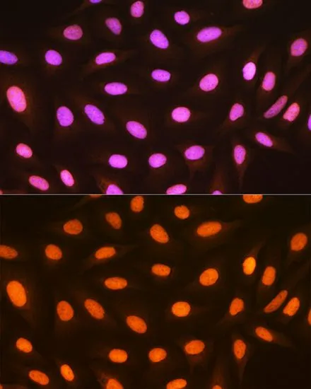
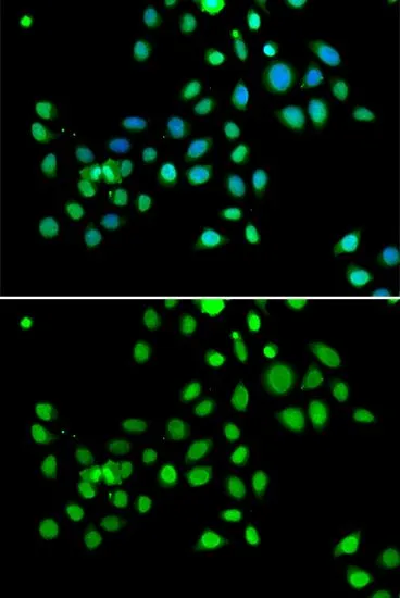
ICC/IF analysis of U2OS cells using GTX03241 Macro H2A.1 antibody [GT1329]. Blue : DAPI for nuclear staining Dilution : 1:100
![IHC-P analysis of mouse lung tissue section using GTX03241 Macro H2A.1 antibody [GT1329]. Dilution : 1:100](https://www.genetex.com/upload/website/prouct_img/normal/GTX03241/GTX03241_20210615_IHC-P_58_w_23053123_417.webp "IHC-P analysis of mouse lung tissue section using GTX03241 Macro H2A.1 antibody [GT1329]. Dilution : 1:100")
![IHC-P analysis of rat brain tissue section using GTX03241 Macro H2A.1 antibody [GT1329]. Dilution : 1:100](https://www.genetex.com/upload/website/prouct_img/normal/GTX03241/GTX03241_20210615_IHC-P_56_w_23053123_294.webp "IHC-P analysis of rat brain tissue section using GTX03241 Macro H2A.1 antibody [GT1329]. Dilution : 1:100")
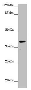
![Whole cell extract (30 μg) was separated by 10% SDS-PAGE, and the membrane was blotted with Macro H2A.1 antibody [GT1329] (GTX03241) diluted at 1:5000. The HRP-conjugated anti-rabbit IgG antibody (GTX213110-01) was used to detect the primary antibody.](https://www.genetex.com/upload/website/prouct_img/normal/GTX03241/GTX03241_4000001396_20210716_WB_2_w_23053123_861.webp "Whole cell extract (30 μg) was separated by 10% SDS-PAGE, and the membrane was blotted with Macro H2A.1 antibody [GT1329] (GTX03241) diluted at 1:5000. The HRP-conjugated anti-rabbit IgG antibody (GTX213110-01) was used to detect the primary antibody.")
![ICC/IF analysis of C6 cells using GTX03241 Macro H2A.1 antibody [GT1329]. Blue : DAPI for nuclear staining Dilution : 1:100](https://www.genetex.com/upload/website/prouct_img/normal/GTX03241/GTX03241_20210615_ICCIF_59_w_23053123_451.webp "ICC/IF analysis of C6 cells using GTX03241 Macro H2A.1 antibody [GT1329]. Blue : DAPI for nuclear staining Dilution : 1:100")
![IHC-P analysis of human liver cancer section using GTX03241 Macro H2A.1 antibody [GT1329]. Dilution : 1:100](https://www.genetex.com/upload/website/prouct_img/normal/GTX03241/GTX03241_20210615_IHC-P_57_w_23053123_308.webp "IHC-P analysis of human liver cancer section using GTX03241 Macro H2A.1 antibody [GT1329]. Dilution : 1:100")
![Whole cell extract (30 μg) was separated by 10% SDS-PAGE, and the membrane was blotted with Macro H2A.1 antibody [GT1329] (GTX03241) diluted at 1:5000. The HRP-conjugated anti-rabbit IgG antibody (GTX213110-01) was used to detect the primary antibody.](https://www.genetex.com/upload/website/prouct_img/normal/GTX03241/GTX03241_4000001396_20210716_WB_w_23053123_831.webp "Whole cell extract (30 μg) was separated by 10% SDS-PAGE, and the membrane was blotted with Macro H2A.1 antibody [GT1329] (GTX03241) diluted at 1:5000. The HRP-conjugated anti-rabbit IgG antibody (GTX213110-01) was used to detect the primary antibody.")
![WB analysis of various samples using GTX03241 Macro H2A.1 antibody [GT1329]. Dilution : 1:1000 Loading : 25μg per lane](https://www.genetex.com/upload/website/prouct_img/normal/GTX03241/GTX03241_54_WB_w_23053123_801.webp "WB analysis of various samples using GTX03241 Macro H2A.1 antibody [GT1329]. Dilution : 1:1000 Loading : 25μg per lane")
ICC/IF analysis of U2OS cells using GTX03241 Macro H2A.1 antibody [GT1329]. Blue : DAPI for nuclear staining Dilution : 1:100
Macro H2A.1 antibody [GT1329]
GTX03241
ApplicationsImmunoFluorescence, Western Blot, ImmunoCytoChemistry, ImmunoHistoChemistry, ImmunoHistoChemistry Paraffin
Product group Antibodies
ReactivityHuman, Mouse, Rat
TargetMACROH2A1
Overview
- SupplierGeneTex
- Product NameMacro H2A.1 antibody [GT1329]
- Delivery Days Customer9
- Application Supplier NoteWB: 1:500 - 1:2000. ICC/IF: 1:50 - 1:200. IHC-P: 1:50 - 1:200. *Optimal dilutions/concentrations should be determined by the researcher.Not tested in other applications.
- ApplicationsImmunoFluorescence, Western Blot, ImmunoCytoChemistry, ImmunoHistoChemistry, ImmunoHistoChemistry Paraffin
- CertificationResearch Use Only
- ClonalityMonoclonal
- Clone IDGT1329
- Concentration0.95 mg/ml
- ConjugateUnconjugated
- Gene ID9555
- Target nameMACROH2A1
- Target descriptionmacroH2A.1 histone
- Target synonymsH2A.y, H2A/y, H2AF12M, H2AFY, MACROH2A1.1, mH2A1, macroH2A1.2, core histone macro-H2A.1, H2A histone family member Y, histone H2A.y, histone macroH2A1, histone macroH2A1.1, histone macroH2A1.2, medulloblastoma antigen MU-MB-50.205
- HostRabbit
- IsotypeIgG
- Protein IDO75367
- Protein NameCore histone macro-H2A.1
- Scientific DescriptionHistones are basic nuclear proteins that are responsible for the nucleosome structure of the chromosomal fiber in eukaryotes. Nucleosomes consist of approximately 146 bp of DNA wrapped around a histone octamer composed of pairs of each of the four core histones (H2A, H2B, H3, and H4). The chromatin fiber is further compacted through the interaction of a linker histone, H1, with the DNA between the nucleosomes to form higher order chromatin structures. This gene encodes a replication-independent histone that is a member of the histone H2A family. It replaces conventional H2A histones in a subset of nucleosomes where it represses transcription and participates in stable X chromosome inactivation. Alternative splicing results in multiple transcript variants encoding different isoforms. [provided by RefSeq, Oct 2015]
- ReactivityHuman, Mouse, Rat
- Storage Instruction-20°C or -80°C,2°C to 8°C
- UNSPSC41116161
Datasheet
Related products

Product group Antibodies
Anti-H2AFY AntibodyAMAB91347
ApplicationsWestern Blot, ImmunoCytoChemistry, ImmunoHistoChemistry
ReactivityHuman, Mouse, Rat
TargetMACROH2A1
- SizePrice
Product group Antibodies
Anti-H2AFY AntibodyA31835
ApplicationsImmunoFluorescence, Western Blot, ImmunoHistoChemistry
ReactivityHuman, Mouse, Rat
- SizePrice
Product group Antibodies
H2AFY AntibodyCSB-PA010098DSR1HU
ApplicationsWestern Blot, ELISA, ImmunoHistoChemistry
ReactivityHuman
TargetMACROH2A1
- SizePrice

Product group Antibodies
Anti-H2AFY/MACROH2A1 Antibody Picoband(r)A04635-3-CARRIER-FREE
ApplicationsImmunoFluorescence, Western Blot, ELISA, ImmunoCytoChemistry, ImmunoHistoChemistry
ReactivityHuman, Mouse, Rat
TargetMACROH2A1
- SizePrice
Product group Antibodies

H2AFY / MACROH2A1 AntibodyLS-C482756
ApplicationsImmunoFluorescence, Western Blot, ImmunoCytoChemistry, ImmunoHistoChemistry, ImmunoHistoChemistry Paraffin
ReactivityHuman, Mouse, Rat
TargetMACROH2A1
- SizePrice
Product group Antibodies
macro H2A.1 Recombinant Antibody, AbBy Fluor-594 ConjugatedBSM-61665R-BF594
ApplicationsImmunoFluorescence, Western Blot
ReactivityHuman, Mouse, Rat
TargetMACROH2A1
- SizePrice
Product group Antibodies
ApplicationsWestern Blot, ImmunoHistoChemistry
ReactivityPorcine
TargetMACROH2A1
- SizePrice
Product group Antibodies
Macro H2A.1 antibodyGTX32709
ApplicationsImmunoFluorescence, Western Blot, ImmunoCytoChemistry, ImmunoHistoChemistry, ImmunoHistoChemistry Paraffin
ReactivityHuman, Mouse, Rat
TargetMACROH2A1
- SizePrice
Product group Antibodies
Anti-H2AFY Antibody144-07045
ApplicationsImmunoFluorescence, Western Blot, ImmunoHistoChemistry
ReactivityHuman, Mouse, Rat
TargetMACROH2A1
- SizePrice