


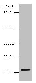
Figure 1. Western blot analysis of HDDC3 using anti-HDDC3 antibody (A12287-1). Electrophoresis was performed on a 5-20% SDS-PAGE gel at 70V (Stacking gel) / 90V (Resolving gel) for 2-3 hours. The sample well of each lane was loaded with 30 ug of sample under reducing conditions. Lane 1: mouse heart tissue lysates. After electrophoresis, proteins were transferred to a nitrocellulose membrane at 150 mA for 50-90 minutes. Blocked the membrane with 5% non-fat milk/TBS for 1.5 hour at RT. The membrane was incubated with rabbit anti-HDDC3 antigen affinity purified polyclonal antibody (Catalog # A12287-1) at 0.5 microg/mL overnight at 4°C, then washed with TBS-0.1%Tween 3 times with 5 minutes each and probed with a goat anti-rabbit IgG-HRP secondary antibody at a dilution of 1:5000 for 1.5 hour at RT. The signal is developed using an Enhanced Chemiluminescent detection (ECL) kit (Catalog # EK1002) with Tanon 5200 system. A specific band was detected for HDDC3 at approximately 22 kDa. The expected band size for HDDC3 is at 20 kDa.
Figure 1. Western blot analysis of HDDC3 using anti-HDDC3 antibody (A12287-1). Electrophoresis was performed on a 5-20% SDS-PAGE gel at 70V (Stacking gel) / 90V (Resolving gel) for 2-3 hours. The sample well of each lane was loaded with 30 ug of sample under reducing conditions. Lane 1: mouse heart tissue lysates. After electrophoresis, proteins were transferred to a nitrocellulose membrane at 150 mA for 50-90 minutes. Blocked the membrane with 5% non-fat milk/TBS for 1.5 hour at RT. The membrane was incubated with rabbit anti-HDDC3 antigen affinity purified polyclonal antibody (Catalog # A12287-1) at 0.5 microg/mL overnight at 4°C, then washed with TBS-0.1%Tween 3 times with 5 minutes each and probed with a goat anti-rabbit IgG-HRP secondary antibody at a dilution of 1:5000 for 1.5 hour at RT. The signal is developed using an Enhanced Chemiluminescent detection (ECL) kit (Catalog # EK1002) with Tanon 5200 system. A specific band was detected for HDDC3 at approximately 22 kDa. The expected band size for HDDC3 is at 20 kDa.
Anti-HDDC3 Antibody Picoband(r)
A12287-1-CARRIER-FREE
ApplicationsWestern Blot, ELISA
Product group Antibodies
ReactivityHuman, Mouse
TargetHDDC3
Overview
- SupplierBoster Bio
- Product NameAnti-HDDC3 Antibody Picoband(r)
- Delivery Days Customer9
- ApplicationsWestern Blot, ELISA
- CertificationResearch Use Only
- ClonalityPolyclonal
- Concentration500 ug/ml
- Gene ID374659
- Target nameHDDC3
- Target descriptionHD domain containing 3
- Target synonyms(ppGpp)ase, MESH1, MYNRL15, guanosine-3',5'-bis(diphosphate) 3'-pyrophosphohydrolase MESH1, HD domain-containing protein 3, metazoan SpoT homolog 1, myeloid leukemia noncoding regulatory locus on chromosome 15, penta-phosphate guanosine-3'-pyrophosphohydrolase
- HostRabbit
- Protein IDQ8N4P3
- Protein NameGuanosine-3',5'-bis(diphosphate) 3'-pyrophosphohydrolase MESH1
- Scientific DescriptionBoster Bio Anti-HDDC3 Antibody Picoband® catalog # A12287-1. Tested in WB, ELISA applications. This antibody reacts with Human, Mouse. The brand Picoband indicates this is a premium antibody that guarantees superior quality, high affinity, and strong signals with minimal background in Western blot applications. Only our best-performing antibodies are designated as Picoband, ensuring unmatched performance.
- ReactivityHuman, Mouse
- Storage Instruction-20°C,2°C to 8°C
- UNSPSC12352203
Related products
Product group Antibodies
HDDC3 Polyclonal AntibodyBS-8061R
ApplicationsImmunoFluorescence, Western Blot, ELISA, ImmunoCytoChemistry, ImmunoHistoChemistry, ImmunoHistoChemistry Frozen, ImmunoHistoChemistry Paraffin
ReactivityBovine, Canine, Equine, Human, Mouse, Rabbit, Rat
TargetHDDC3
- SizePrice
Product group Antibodies
HDDC3 AntibodyCSB-PA836666HA01HU
ApplicationsWestern Blot, ELISA, ImmunoHistoChemistry
ReactivityHuman, Mouse
TargetHDDC3
- SizePrice
Product group Antibodies
Hddc3 Polyclonal AntibodyCAC09533
ApplicationsWestern Blot, ELISA, ImmunoHistoChemistry
ReactivityMouse
TargetHDDC3
- SizePrice
Product group Antibodies

HDDC3 Antibody (aa2-140)LS-C371727
ApplicationsWestern Blot, ELISA, ImmunoHistoChemistry, ImmunoHistoChemistry Paraffin
ReactivityHuman
TargetHDDC3
- SizePrice
Product group Antibodies
Anti-HDDC3 AntibodyHPA040895
ApplicationsWestern Blot, ImmunoCytoChemistry, ImmunoHistoChemistry
ReactivityHuman
TargetHDDC3
- SizePrice
Product group Antibodies
HDDC3 antibody, InternalGTX45162
ApplicationsWestern Blot
ReactivityHuman
TargetHDDC3
- SizePrice
Product group Antibodies
HDDC3 AntibodyPACO38538
ApplicationsWestern Blot, ELISA, ImmunoHistoChemistry
ReactivityHuman, Mouse
TargetHDDC3
- SizePrice