


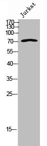
Figure 1. Western blot analysis of NDC80 using anti-NDC80 antibody (A01731-2). Electrophoresis was performed on a 5-20% SDS-PAGE gel at 70V (Stacking gel) / 90V (Resolving gel) for 2-3 hours. The sample well of each lane was loaded with 50ug of sample under reducing conditions. Lane 1: human K562 whole cell lysates, Lane 2: rat spleen tissue lysates, Lane 3: mouse spleen tissue lysates. After Electrophoresis, proteins were transferred to a Nitrocellulose membrane at 150mA for 50-90 minutes. Blocked the membrane with 5% Non-fat Milk/ TBS for 1.5 hour at RT. The membrane was incubated with rabbit anti-NDC80 antigen affinity purified polyclonal antibody (Catalog # A01731-2) at 0.5 microg/mL overnight at 4°C, then washed with TBS-0.1%Tween 3 times with 5 minutes each and probed with a goat anti-rabbit IgG-HRP secondary antibody at a dilution of 1:10000 for 1.5 hour at RT. The signal is developed using an Enhanced Chemiluminescent detection (ECL) kit (Catalog # EK1002) with Tanon 5200 system. A specific band was detected for NDC80 at approximately 74KD. The expected band size for NDC80 is at 74KD.
. NDC80 was detected in immunocytochemical section of U2OS cells. Enzyme antigen retrieval was performed using IHC enzyme antigen retrieval reagent (AR0022) for 15 mins. The cells were blocked with 10% goat serum. And then incubated with 2microg/mL rabbit anti-NDC80 Antibody (A01731-2) overnight at 4°C. DyLight®488 Conjugated Goat Anti-Rabbit IgG (BA1127) was used as secondary antibody at 1:100 dilution and incubated for 30 minutes at 37°C. The section was counterstained with DAPI. Visualize using a fluorescence microscope and filter sets appropriate for the label used.")
. Overlay histogram showing HL-60 cells stained with A01731-2 (Blue line). To facilitate intracellular staining, cells were fixed with 4% paraformaldehyde and permeabilized with permeabilization buffer. The cells were blocked with 10% normal goat serum. And then incubated with rabbit anti-NDC80 Antibody (A01731-2, 1microg/1x106 cells) for 30 min at 20°C. DyLight®488 conjugated goat anti-rabbit IgG (BA1127, 5-10microg/1x106 cells) was used as secondary antibody for 30 minutes at 20°C. Isotype control antibody (Green line) was rabbit IgG (1microg/1x106) used under the same conditions. Unlabelled sample without incubation with primary antibody and secondary antibody (Red line) was used as a blank control.")
. Overlay histogram showing SiHa cells stained with A01731-2 (Blue line). To facilitate intracellular staining, cells were fixed with 4% paraformaldehyde and permeabilized with permeabilization buffer. The cells were blocked with 10% normal goat serum. And then incubated with rabbit anti-NDC80 Antibody (A01731-2, 1microg/1x106 cells) for 30 min at 20°C. DyLight®488 conjugated goat anti-rabbit IgG (BA1127, 5-10microg/1x106 cells) was used as secondary antibody for 30 minutes at 20°C. Isotype control antibody (Green line) was rabbit IgG (1microg/1x106) used under the same conditions. Unlabelled sample without incubation with primary antibody and secondary antibody (Red line) was used as a blank control.")
. NDC80 was detected in immunocytochemical section of U20S cells. Enzyme antigen retrieval was performed using IHC enzyme antigen retrieval reagent (AR0022) for 15 mins. The cells were blocked with 10% goat serum. And then incubated with 2microg/mL rabbit anti-NDC80 Antibody (A01731-2) overnight at 4°C. DyLight®594 Conjugated Goat Anti-Rabbit IgG (BA1142) was used as secondary antibody at 1:100 dilution and incubated for 30 minutes at 37°C. The section was counterstained with DAPI. Visualize using a fluorescence microscope and filter sets appropriate for the label used.")
. NDC80 was detected in immunocytochemical section of U20S cells. Enzyme antigen retrieval was performed using IHC enzyme antigen retrieval reagent (AR0022) for 15 mins. The cells were blocked with 10% goat serum. And then incubated with 2microg/mL rabbit anti-NDC80 Antibody (A01731-2) overnight at 4°C. DyLight®488 Conjugated Goat Anti-Rabbit IgG (BA1127) was used as secondary antibody at 1:100 dilution and incubated for 30 minutes at 37°C. The section was counterstained with DAPI. Visualize using a fluorescence microscope and filter sets appropriate for the label used.")
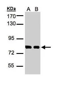
Figure 1. Western blot analysis of NDC80 using anti-NDC80 antibody (A01731-2). Electrophoresis was performed on a 5-20% SDS-PAGE gel at 70V (Stacking gel) / 90V (Resolving gel) for 2-3 hours. The sample well of each lane was loaded with 50ug of sample under reducing conditions. Lane 1: human K562 whole cell lysates, Lane 2: rat spleen tissue lysates, Lane 3: mouse spleen tissue lysates. After Electrophoresis, proteins were transferred to a Nitrocellulose membrane at 150mA for 50-90 minutes. Blocked the membrane with 5% Non-fat Milk/ TBS for 1.5 hour at RT. The membrane was incubated with rabbit anti-NDC80 antigen affinity purified polyclonal antibody (Catalog # A01731-2) at 0.5 microg/mL overnight at 4°C, then washed with TBS-0.1%Tween 3 times with 5 minutes each and probed with a goat anti-rabbit IgG-HRP secondary antibody at a dilution of 1:10000 for 1.5 hour at RT. The signal is developed using an Enhanced Chemiluminescent detection (ECL) kit (Catalog # EK1002) with Tanon 5200 system. A specific band was detected for NDC80 at approximately 74KD. The expected band size for NDC80 is at 74KD.
Anti-HEC1/HEC/NDC80 Antibody Picoband(r)
A01731-2-CARRIER-FREE
ApplicationsFlow Cytometry, ImmunoFluorescence, Western Blot, ELISA, ImmunoCytoChemistry
Product group Antibodies
ReactivityHuman, Mouse, Rat
TargetNDC80
Overview
- SupplierBoster Bio
- Product NameAnti-HEC1/HEC/NDC80 Antibody Picoband(r)
- Delivery Days Customer9
- ApplicationsFlow Cytometry, ImmunoFluorescence, Western Blot, ELISA, ImmunoCytoChemistry
- CertificationResearch Use Only
- ClonalityPolyclonal
- Concentration500 ug/ml
- Gene ID10403
- Target nameNDC80
- Target descriptionNDC80 kinetochore complex component
- Target synonymsHEC, HEC1, HsHec1, KNTC2, TID3, hsNDC80, kinetochore protein NDC80 homolog, NDC80 homolog, kinetochore complex component, NDC80 kinetochore complex component homolog, highly expressed in cancer protein, highly expressed in cancer, rich in leucine heptad repeats, kinetochore associated 2, kinetochore protein Hec1, kinetochore-associated protein 2, retinoblastoma-associated protein HEC
- HostRabbit
- IsotypeIgG
- Protein IDO14777
- Protein NameKinetochore protein NDC80 homolog
- Scientific DescriptionBoster Bio Anti-HEC1/HEC/NDC80 Antibody Picoband® catalog # A01731-2. Tested in ELISA, Flow Cytometry, IF, ICC, WB applications. This antibody reacts with Human, Mouse, Rat. The brand Picoband indicates this is a premium antibody that guarantees superior quality, high affinity, and strong signals with minimal background in Western blot applications. Only our best-performing antibodies are designated as Picoband, ensuring unmatched performance.
- ReactivityHuman, Mouse, Rat
- Storage Instruction-20°C,2°C to 8°C
- UNSPSC12352203
Related products

Product group Antibodies
Anti-KNTC2 AntibodyA97839
ApplicationsImmunoFluorescence, Western Blot, ELISA
ReactivityHuman, Mouse
- SizePrice
Product group Antibodies

Anti-Kinetochore protein NDC80 homolog [Hec1 (9G3)]AB03500-1.1-BT
ApplicationsImmunoFluorescence, ImmunoPrecipitation, Western Blot
ReactivityHuman
TargetNDC80
- SizePrice
Product group Antibodies
Anti-NDC80 Antibody144-05411
ApplicationsWestern Blot
ReactivityHuman, Mouse
TargetNDC80
- SizePrice
Product group Antibodies

HEC1 / NDC80 AntibodyLS-C749115
ApplicationsWestern Blot
ReactivityHuman, Mouse, Rat
TargetNDC80
- SizePrice
Product group Antibodies
HEC1 Recombinant Antibody, AbBy Fluor-488 ConjugatedBSM-61551R-BF488
ApplicationsWestern Blot
ReactivityHuman, Mouse, Rat
TargetNDC80
- SizePrice
Product group Antibodies
NDC80 AntibodyCSB-PA006498
ApplicationsImmunoFluorescence, Western Blot, ELISA
ReactivityHuman, Mouse
TargetNDC80
- SizePrice
Product group Antibodies

Goat anti-HEC1EB05610
ApplicationsWestern Blot, ELISA, ImmunoHistoChemistry
ReactivityHuman
TargetNDC80
- SizePrice

Product group Antibodies
Anti-NDC80 AntibodyHPA066330
ApplicationsWestern Blot, ImmunoCytoChemistry
ReactivityHuman
TargetNDC80
- SizePrice
Product group Antibodies
Hec1 antibodyGTX110735
ApplicationsImmunoPrecipitation, Western Blot
ReactivityHuman
TargetNDC80
- SizePrice