


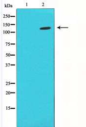
Figure 1. Western blot analysis of HIF1A using anti-HIF1A antibody (A00013-1). Electrophoresis was performed on a 5-20% SDS-PAGE gel at 70V (Stacking gel) / 90V (Resolving gel) for 2-3 hours. The sample well of each lane was loaded with 50ug of sample under reducing conditions. Lane 1: human U-87MG whole cell lysates, Lane 2: human U2OS whole cell lysates, Lane 3: human PC-3 whole cell lysates, Lane 4: human A549 whole cell lysates, Lane 5: human HepG2 whole cell lysates. After Electrophoresis, proteins were transferred to a Nitrocellulose membrane at 150mA for 50-90 minutes. Blocked the membrane with 5% Non-fat Milk/ TBS for 1.5 hour at RT. The membrane was incubated with rabbit anti-HIF1A antigen affinity purified polyclonal antibody (Catalog # A00013-1) at 0.5 microg/mL overnight at 4°C, then washed with TBS-0.1%Tween 3 times with 5 minutes each and probed with a goat anti-rabbit IgG-HRP secondary antibody at a dilution of 1:10000 for 1.5 hour at RT. The signal is developed using an Enhanced Chemiluminescent detection (ECL) kit (Catalog # EK1002) with Tanon 5200 system. A specific band was detected for HIF1A at approximately 123KD. The expected band size for HIF1A is at 93KD.
Figure 1. Western blot analysis of HIF1A using anti-HIF1A antibody (A00013-1). Electrophoresis was performed on a 5-20% SDS-PAGE gel at 70V (Stacking gel) / 90V (Resolving gel) for 2-3 hours. The sample well of each lane was loaded with 50ug of sample under reducing conditions. Lane 1: human U-87MG whole cell lysates, Lane 2: human U2OS whole cell lysates, Lane 3: human PC-3 whole cell lysates, Lane 4: human A549 whole cell lysates, Lane 5: human HepG2 whole cell lysates. After Electrophoresis, proteins were transferred to a Nitrocellulose membrane at 150mA for 50-90 minutes. Blocked the membrane with 5% Non-fat Milk/ TBS for 1.5 hour at RT. The membrane was incubated with rabbit anti-HIF1A antigen affinity purified polyclonal antibody (Catalog # A00013-1) at 0.5 microg/mL overnight at 4°C, then washed with TBS-0.1%Tween 3 times with 5 minutes each and probed with a goat anti-rabbit IgG-HRP secondary antibody at a dilution of 1:10000 for 1.5 hour at RT. The signal is developed using an Enhanced Chemiluminescent detection (ECL) kit (Catalog # EK1002) with Tanon 5200 system. A specific band was detected for HIF1A at approximately 123KD. The expected band size for HIF1A is at 93KD.
Anti-HIF-1 alpha/HIF1A Antibody Picoband(r)
A00013-1-CARRIER-FREE
ApplicationsWestern Blot, ELISA
Product group Antibodies
ReactivityHuman
TargetHIF1A
Overview
- SupplierBoster Bio
- Product NameAnti-HIF-1 alpha/HIF1A Antibody Picoband(r)
- Delivery Days Customer9
- ApplicationsWestern Blot, ELISA
- CertificationResearch Use Only
- ClonalityPolyclonal
- Concentration500 ug/ml
- Gene ID3091
- Target nameHIF1A
- Target descriptionhypoxia inducible factor 1 subunit alpha
- Target synonymsHIF-1-alpha, HIF-1A, HIF-1alpha, HIF1, HIF1-ALPHA, MOP1, PASD8, bHLHe78, hypoxia-inducible factor 1-alpha, ARNT interacting protein, PAS domain-containing protein 8, basic-helix-loop-helix-PAS protein MOP1, class E basic helix-loop-helix protein 78, hypoxia inducible factor 1 alpha subunit, hypoxia inducible factor 1, alpha subunit (basic helix-loop-helix transcription factor), hypoxia-inducible factor1alpha, member of PAS protein 1, member of PAS superfamily 1
- HostRabbit
- IsotypeIgG
- Protein IDQ16665
- Protein NameHypoxia-inducible factor 1-alpha
- Scientific DescriptionBoster Bio Anti-HIF-1 alpha/HIF1A Antibody Picoband® catalog # A00013-1. Tested in ELISA, WB applications. This antibody reacts with Human. The brand Picoband indicates this is a premium antibody that guarantees superior quality, high affinity, and strong signals with minimal background in Western blot applications. Only our best-performing antibodies are designated as Picoband, ensuring unmatched performance.
- ReactivityHuman
- Storage Instruction-20°C,2°C to 8°C
- UNSPSC12352203
Related products
Product group Antibodies
Anti-HIF1A AntibodyA37506
ApplicationsWestern Blot, ImmunoHistoChemistry
ReactivityHuman, Mouse, Rat
- SizePrice
Product group Antibodies
anti-Hif-1 alpha (human), mAb (ANC10G3)ANC-335-020
ApplicationsFlow Cytometry, ELISA
ReactivityHuman
TargetHIF1A
- SizePrice
Product group Antibodies

HIF1A / HIF1 Alpha AntibodyLS-C831817
ApplicationsELISA, ImmunoHistoChemistry
ReactivityHuman, Mouse
TargetHIF1A
- SizePrice
Product group Antibodies
References
HIF-1 Alpha Polyclonal AntibodyBS-0737R
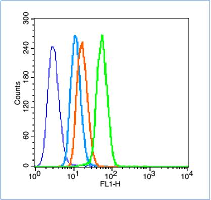
ApplicationsFlow Cytometry, ImmunoFluorescence, ImmunoPrecipitation, Western Blot, ELISA, ImmunoCytoChemistry, ImmunoHistoChemistry, ImmunoHistoChemistry Frozen, ImmunoHistoChemistry Paraffin
ReactivityChicken, Human, Mouse, Rat
TargetHIF1A
- SizePrice
Product group Antibodies
HIF1A AntibodyCSB-PA002906
ApplicationsWestern Blot, ELISA, ImmunoHistoChemistry
ReactivityHuman, Mouse, Rat
TargetHIF1A
- SizePrice
Product group Antibodies
Hif1A Polyclonal AntibodyCAC07003
ApplicationsImmunoFluorescence, ELISA, ImmunoHistoChemistry
TargetHIF1A
- SizePrice

Product group Antibodies
Anti-HIF1A AntibodyHPA000907
ApplicationsImmunoCytoChemistry
ReactivityHuman
TargetHIF1A
- SizePrice
Product group Antibodies
HIF1 alpha antibodyGTX127309
ApplicationsImmunoFluorescence, ImmunoPrecipitation, Western Blot, ChIP Chromatin ImmunoPrecipitation, ImmunoCytoChemistry, ImmunoHistoChemistry, ImmunoHistoChemistry Frozen, ImmunoHistoChemistry Paraffin
ReactivityBovine, Human, Mouse, Rabbit, Rat
TargetHIF1A
- SizePrice