


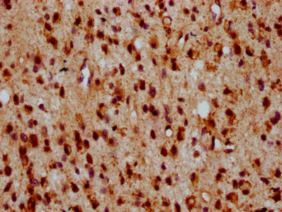
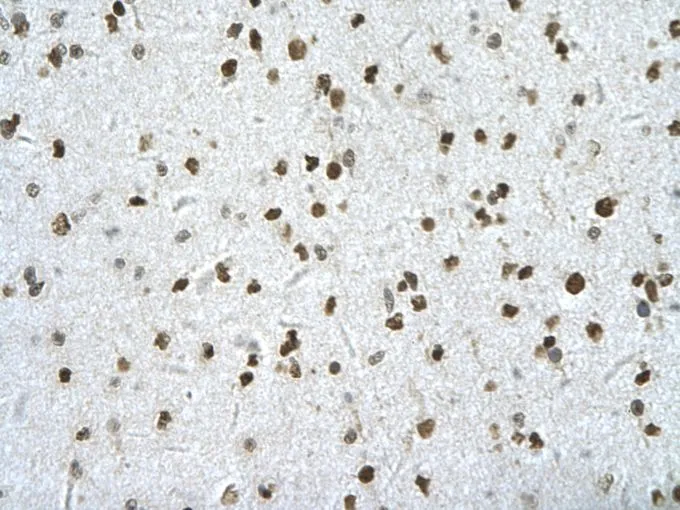
Immunohistochemical staining of human skeletal muscle shows strong cytoplasmic positivity in myocytes.
Immunohistochemical staining of human skeletal muscle shows strong cytoplasmic positivity in myocytes.
Anti-HIPK2 Antibody
HPA007611
ApplicationsImmunoHistoChemistry
Product group Antibodies
ReactivityHuman
TargetHIPK2
Overview
- SupplierAtlas Antibodies
- Product NameAnti-HIPK2 Antibody
- Delivery Days Customer4
- ApplicationsImmunoHistoChemistry
- CertificationResearch Use Only
- ClonalityPolyclonal
- ConjugateUnconjugated
- Gene ID28996
- Target nameHIPK2
- Target descriptionhomeodomain interacting protein kinase 2
- Target synonymsPRO0593, homeodomain-interacting protein kinase 2, hHIPk2
- HostRabbit
- IsotypeIgG
- Protein IDQ9H2X6
- Protein NameHomeodomain-interacting protein kinase 2
- Scientific DescriptionRecombinant Protein Epitope Signature Tag (PrEST) antigen sequence
- ReactivityHuman
- Storage Instruction-20°C,2°C to 8°C
- UNSPSC41116161
Datasheet
MSDS
Related products

Product group Antibodies
Anti-HIPK2 AntibodyA28507
ApplicationsWestern Blot
ReactivityHuman, Mouse, Rat
- SizePrice

Product group Antibodies
ApplicationsFlow Cytometry, ImmunoFluorescence, ImmunoPrecipitation, Western Blot, ImmunoCytoChemistry
ReactivityHuman, Mouse
TargetHIPK2
- SizePrice
Product group Antibodies

HIPK2 AntibodyLS-C830005
ApplicationsELISA, ImmunoHistoChemistry
ReactivityHuman, Mouse
TargetHIPK2
- SizePrice
Product group Antibodies
HIPK2 AntibodyCSB-PA867138LA01HU
ApplicationsELISA, ImmunoHistoChemistry
ReactivityHuman
TargetHIPK2
- SizePrice
Product group Antibodies
HIPK2 antibody, InternalGTX30803
ApplicationsWestern Blot, ImmunoHistoChemistry, ImmunoHistoChemistry Paraffin
ReactivityHuman
TargetHIPK2
- SizePrice
Product group Antibodies
ApplicationsImmunoPrecipitation, Western Blot, ImmunoCytoChemistry, ImmunoHistoChemistry
ReactivityMouse, Rat
TargetHIPK2
- SizePrice
Product group Antibodies
HIPK2 Recombinant Antibody, AbBy Fluor-555 ConjugatedBSM-62009R-BF555
ApplicationsFlow Cytometry, Western Blot
ReactivityHuman, Mouse
TargetHIPK2
- SizePrice