

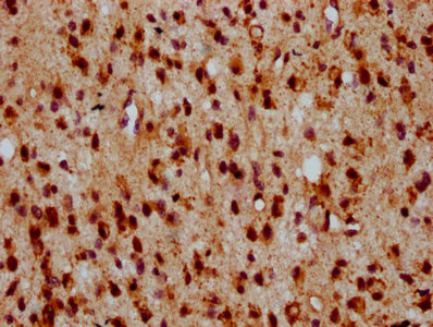
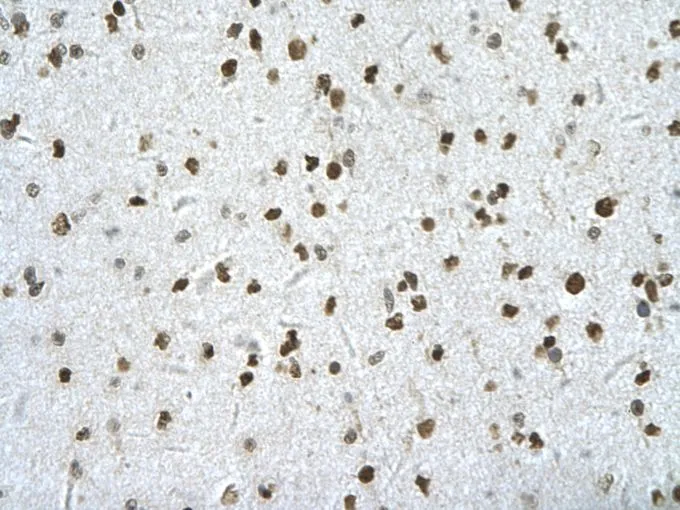
IHC image of CSB-PA867138LA01HU diluted at 1:500 and staining in paraffin-embedded human glioma performed on a Leica BondTM system. After dewaxing and hydration, antigen retrieval was mediated by high pressure in a citrate buffer (pH 6.0). Section was blocked with 10% normal goat serum 30min at RT. Then primary antibody (1% BSA) was incubated at 4°C overnight. The primary is detected by a biotinylated secondary antibody and visualized using an HRP conjugated SP system.
. Section was blocked with 10% normal goat serum 30min at RT. Then primary antibody (1% BSA) was incubated at 4°C overnight. The primary is detected by a biotinylated secondary antibody and visualized using an HRP conjugated SP system.")
IHC image of CSB-PA867138LA01HU diluted at 1:500 and staining in paraffin-embedded human glioma performed on a Leica BondTM system. After dewaxing and hydration, antigen retrieval was mediated by high pressure in a citrate buffer (pH 6.0). Section was blocked with 10% normal goat serum 30min at RT. Then primary antibody (1% BSA) was incubated at 4°C overnight. The primary is detected by a biotinylated secondary antibody and visualized using an HRP conjugated SP system.
HIPK2 Antibody
CSB-PA867138LA01HU
ApplicationsELISA, ImmunoHistoChemistry
Product group Antibodies
ReactivityHuman
TargetHIPK2
Overview
- SupplierCusabio
- Product NameHIPK2 Antibody
- Delivery Days Customer20
- ApplicationsELISA, ImmunoHistoChemistry
- CertificationResearch Use Only
- ClonalityPolyclonal
- ConjugateUnconjugated
- Gene ID28996
- Target nameHIPK2
- Target descriptionhomeodomain interacting protein kinase 2
- Target synonymsPRO0593, homeodomain-interacting protein kinase 2, hHIPk2
- HostRabbit
- IsotypeIgG
- Protein IDQ9H2X6
- Protein NameHomeodomain-interacting protein kinase 2
- Scientific DescriptionSerine/threonine-protein kinase involved in transcription regulation, p53/TP53-mediated cellular apoptosis and regulation of the cell cycle. Acts as a corepressor of several transcription factors, including SMAD1 and POU4F1/Brn3a and probably NK homeodomain transcription factors. Phosphorylates PDX1, ATF1, PML, p53/TP53, CREB1, CTBP1, CBX4, RUNX1, EP300, CTNNB1, HMGA1 and ZBTB4. Inhibits cell growth and promotes apoptosis through the activation of p53/TP53 both at the transcription level and at the protein level (by phosphorylation and indirect acetylation). The phosphorylation of p53/TP53 may be mediated by a p53/TP53-HIPK2-AXIN1 complex. Involved in the response to hypoxia by acting as a transcriptional co-suppressor of HIF1A. Mediates transcriptional activation of TP73. In response to TGFB, cooperates with DAXX to activate JNK. Negative regulator through phosphorylation and subsequent proteasomal degradation of CTNNB1 and the antiapoptotic factor CTBP1. In the Wnt/beta-catenin signaling pathway acts as an intermediate kinase between MAP3K7/TAK1 and NLK to promote the proteasomal degradation of MYB. Phosphorylates CBX4 upon DNA damage and promotes its E3 SUMO-protein ligase activity. Activates CREB1 and ATF1 transcription factors by phosphorylation in response to genotoxic stress. In response to DNA damage, stabilizes PML by phosphorylation. PML, HIPK2 and FBXO3 may act synergically to activate p53/TP53-dependent transactivation. Promotes angiogenesis, and is involved in erythroid differentiation, especially during fetal liver erythropoiesis. Phosphorylation of RUNX1 and EP300 stimulates EP300 transcription regulation activity. Triggers ZBTB4 protein degradation in response to DNA damage. Modulates HMGA1 DNA-binding affinity. In response to high glucose, triggers phosphorylation-mediated subnuclear localization shifting of PDX1. Involved in the regulation of eye size, lens formation and retinal lamination during late embryogenesis.
- ReactivityHuman
- Storage Instruction-20°C or -80°C
- UNSPSC41116161
Related products

Product group Antibodies
Anti-HIPK2 AntibodyA28507
ApplicationsWestern Blot
ReactivityHuman, Mouse, Rat
- SizePrice

Product group Antibodies
ApplicationsFlow Cytometry, ImmunoFluorescence, ImmunoPrecipitation, Western Blot, ImmunoCytoChemistry
ReactivityHuman, Mouse
TargetHIPK2
- SizePrice
Product group Antibodies

HIPK2 AntibodyLS-C830005
ApplicationsELISA, ImmunoHistoChemistry
ReactivityHuman, Mouse
TargetHIPK2
- SizePrice

Product group Antibodies
Anti-HIPK2 AntibodyHPA007611
ApplicationsImmunoHistoChemistry
ReactivityHuman
TargetHIPK2
- SizePrice
Product group Antibodies
HIPK2 antibody, InternalGTX30803
ApplicationsWestern Blot, ImmunoHistoChemistry, ImmunoHistoChemistry Paraffin
ReactivityHuman
TargetHIPK2
- SizePrice
Product group Antibodies
ApplicationsImmunoPrecipitation, Western Blot, ImmunoCytoChemistry, ImmunoHistoChemistry
ReactivityMouse, Rat
TargetHIPK2
- SizePrice
Product group Antibodies
HIPK2 Recombinant Antibody, AbBy Fluor-555 ConjugatedBSM-62009R-BF555
ApplicationsFlow Cytometry, Western Blot
ReactivityHuman, Mouse
TargetHIPK2
- SizePrice