


Figure 1. Western blot analysis of HIPK2 using anti-HIPK2 antibody (M01371). Electrophoresis was performed on a 5-20% SDS-PAGE gel at 70V (Stacking gel) / 90V (Resolving gel) for 2-3 hours. The sample well of each lane was loaded with 30 ug of sample under reducing conditions. Lane 1: human HT-1080 whole cell lysates, Lane 2: human Raji whole cell lysates, Lane 3: human PC-3 whole cell lysates, Lane 4: human HEL whole cell lysates. After electrophoresis, proteins were transferred to a nitrocellulose membrane at 150 mA for 50-90 minutes. Blocked the membrane with 5% non-fat milk/TBS for 1.5 hour at RT. The membrane was incubated with rabbit anti-HIPK2 antigen affinity purified monoclonal antibody (Catalog # M01371) at 1:500 overnight at 4°C, then washed with TBS-0.1%Tween 3 times with 5 minutes each and probed with a goat anti-rabbit IgG-HRP secondary antibody at a dilution of 1:500 for 1.5 hour at RT. The signal is developed using an Enhanced Chemiluminescent detection (ECL) kit (Catalog # EK1002) with Tanon 5200 system. A specific band was detected for HIPK2 at approximately 100 kDa. The expected band size for HIPK2 is at 130 kDa.
Figure 1. Western blot analysis of HIPK2 using anti-HIPK2 antibody (M01371). Electrophoresis was performed on a 5-20% SDS-PAGE gel at 70V (Stacking gel) / 90V (Resolving gel) for 2-3 hours. The sample well of each lane was loaded with 30 ug of sample under reducing conditions. Lane 1: human HT-1080 whole cell lysates, Lane 2: human Raji whole cell lysates, Lane 3: human PC-3 whole cell lysates, Lane 4: human HEL whole cell lysates. After electrophoresis, proteins were transferred to a nitrocellulose membrane at 150 mA for 50-90 minutes. Blocked the membrane with 5% non-fat milk/TBS for 1.5 hour at RT. The membrane was incubated with rabbit anti-HIPK2 antigen affinity purified monoclonal antibody (Catalog # M01371) at 1:500 overnight at 4°C, then washed with TBS-0.1%Tween 3 times with 5 minutes each and probed with a goat anti-rabbit IgG-HRP secondary antibody at a dilution of 1:500 for 1.5 hour at RT. The signal is developed using an Enhanced Chemiluminescent detection (ECL) kit (Catalog # EK1002) with Tanon 5200 system. A specific band was detected for HIPK2 at approximately 100 kDa. The expected band size for HIPK2 is at 130 kDa.
Anti-HIPK2 Rabbit Monoclonal Antibody
M01371
ApplicationsFlow Cytometry, ImmunoFluorescence, ImmunoPrecipitation, Western Blot, ImmunoCytoChemistry
Product group Antibodies
ReactivityHuman, Mouse
TargetHIPK2
Overview
- SupplierBoster Bio
- Product NameAnti-HIPK2 Rabbit Monoclonal Antibody
- Delivery Days Customer9
- ApplicationsFlow Cytometry, ImmunoFluorescence, ImmunoPrecipitation, Western Blot, ImmunoCytoChemistry
- CertificationResearch Use Only
- ClonalityMonoclonal
- Clone ID21H33
- Gene ID28996
- Target nameHIPK2
- Target descriptionhomeodomain interacting protein kinase 2
- Target synonymsPRO0593, homeodomain-interacting protein kinase 2, hHIPk2
- HostRabbit
- IsotypeIgG
- Protein IDQ9H2X6
- Protein NameHomeodomain-interacting protein kinase 2
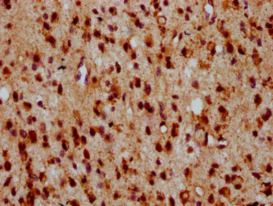
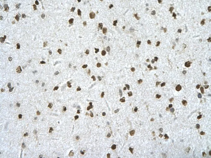
- Scientific DescriptionBoster Bio Anti-HIPK2 Rabbit Monoclonal Antibody catalog # M01371. Tested in WB, ICC/IF, IP, Flow Cytometry applications. This antibody reacts with Human, Mouse.
- ReactivityHuman, Mouse
- Storage Instruction-20°C
- UNSPSC12352203
Related products

Product group Antibodies
Anti-HIPK2 AntibodyA28507
ApplicationsWestern Blot
ReactivityHuman, Mouse, Rat
- SizePrice
Product group Antibodies

HIPK2 AntibodyLS-C830005
ApplicationsELISA, ImmunoHistoChemistry
ReactivityHuman, Mouse
TargetHIPK2
- SizePrice

Product group Antibodies
Anti-HIPK2 AntibodyHPA007611
ApplicationsImmunoHistoChemistry
ReactivityHuman
TargetHIPK2
- SizePrice
Product group Antibodies
HIPK2 AntibodyCSB-PA867138LA01HU
ApplicationsELISA, ImmunoHistoChemistry
ReactivityHuman
TargetHIPK2
- SizePrice
Product group Antibodies
HIPK2 antibody, InternalGTX30803
ApplicationsWestern Blot, ImmunoHistoChemistry, ImmunoHistoChemistry Paraffin
ReactivityHuman
TargetHIPK2
- SizePrice
Product group Antibodies
ApplicationsImmunoPrecipitation, Western Blot, ImmunoCytoChemistry, ImmunoHistoChemistry
ReactivityMouse, Rat
TargetHIPK2
- SizePrice
Product group Antibodies
HIPK2 Recombinant Antibody, AbBy Fluor-555 ConjugatedBSM-62009R-BF555
ApplicationsFlow Cytometry, Western Blot
ReactivityHuman, Mouse
TargetHIPK2
- SizePrice