


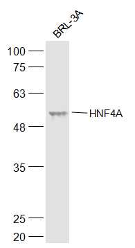
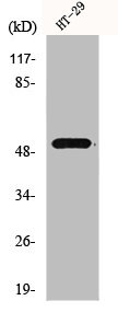
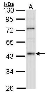
Figure 1. Western blot analysis of HNF-4-alpha using anti-HNF-4-alpha antibody (M00179-1). Electrophoresis was performed on a 5-20% SDS-PAGE gel at 70V (Stacking gel) / 90V (Resolving gel) for 2-3 hours. The sample well of each lane was loaded with 30 ug of sample under reducing conditions. Lane 1: human HepG2 whole cell lysates, Lane 2: rat liver tissue lysates. After electrophoresis, proteins were transferred to a nitrocellulose membrane at 150 mA for 50-90 minutes. Blocked the membrane with 5% non-fat milk/TBS for 1.5 hour at RT. The membrane was incubated with mouse anti-HNF-4-alpha antigen affinity purified monoclonal antibody (Catalog # M00179-1) at 0.5 microg/mL overnight at 4°C, then washed with TBS-0.1%Tween 3 times with 5 minutes each and probed with a goat anti-mouse IgG-HRP secondary antibody at a dilution of 1:10000 for 1.5 hour at RT. The signal is developed using an Enhanced Chemiluminescent detection (ECL) kit (Catalog # EK1001) with Tanon 5200 system. A specific band was detected for HNF-4-alpha at approximately 53 kDa. The expected band size for HNF-4-alpha is at 53 kDa.
. HNF-4-alpha was detected in a paraffin-embedded section of rat colon tissue. Heat mediated antigen retrieval was performed in EDTA buffer (pH 8.0, epitope retrieval solution). The tissue section was blocked with 10% goat serum. The tissue section was then incubated with 2 microg/ml mouse anti-HNF-4-alpha Antibody (M00389-2) overnight at 4°C. Peroxidase Conjugated Goat Anti-mouse IgG was used as secondary antibody and incubated for 30 minutes at 37°C. The tissue section was developed using HRP Conjugated Mouse IgG Super Vision Assay Kit (Catalog # SV0001) with DAB as the chromogen.")
. HNF-4-alpha was detected in a paraffin-embedded section of rat liver tissue. Heat mediated antigen retrieval was performed in EDTA buffer (pH 8.0, epitope retrieval solution). The tissue section was blocked with 10% goat serum. The tissue section was then incubated with 2 microg/ml mouse anti-HNF-4-alpha Antibody (M00389-2) overnight at 4°C. Peroxidase Conjugated Goat Anti-mouse IgG was used as secondary antibody and incubated for 30 minutes at 37°C. The tissue section was developed using HRP Conjugated Mouse IgG Super Vision Assay Kit (Catalog # SV0001) with DAB as the chromogen.")
Figure 1. Western blot analysis of HNF-4-alpha using anti-HNF-4-alpha antibody (M00179-1). Electrophoresis was performed on a 5-20% SDS-PAGE gel at 70V (Stacking gel) / 90V (Resolving gel) for 2-3 hours. The sample well of each lane was loaded with 30 ug of sample under reducing conditions. Lane 1: human HepG2 whole cell lysates, Lane 2: rat liver tissue lysates. After electrophoresis, proteins were transferred to a nitrocellulose membrane at 150 mA for 50-90 minutes. Blocked the membrane with 5% non-fat milk/TBS for 1.5 hour at RT. The membrane was incubated with mouse anti-HNF-4-alpha antigen affinity purified monoclonal antibody (Catalog # M00179-1) at 0.5 microg/mL overnight at 4°C, then washed with TBS-0.1%Tween 3 times with 5 minutes each and probed with a goat anti-mouse IgG-HRP secondary antibody at a dilution of 1:10000 for 1.5 hour at RT. The signal is developed using an Enhanced Chemiluminescent detection (ECL) kit (Catalog # EK1001) with Tanon 5200 system. A specific band was detected for HNF-4-alpha at approximately 53 kDa. The expected band size for HNF-4-alpha is at 53 kDa.
Anti-HNF-4-alpha Antibody Picoband(r) (monoclonal, 6C8E9)
M00389-2-CARRIER-FREE
ApplicationsWestern Blot, ImmunoHistoChemistry
Product group Antibodies
ReactivityHuman, Rat
TargetHNF4A
Overview
- SupplierBoster Bio
- Product NameAnti-HNF-4-alpha Antibody Picoband(r) (monoclonal, 6C8E9)
- Delivery Days Customer9
- ApplicationsWestern Blot, ImmunoHistoChemistry
- CertificationResearch Use Only
- ClonalityMonoclonal
- Clone ID6C8E9
- Concentration500 ug/ml
- Gene ID3172
- Target nameHNF4A
- Target descriptionhepatocyte nuclear factor 4 alpha
- Target synonymsFRTS4, HNF4, HNF4a7, HNF4a8, HNF4a9, HNF4alpha, MODY, MODY1, NR2A1, NR2A21, TCF, TCF-14, TCF14, hepatocyte nuclear factor 4-alpha, HNF4A wild type, HNF4alpha10/11/12, hepatic nuclear factor 4 alpha, nuclear receptor subfamily 2 group A member 1, transcription factor 14, transcription factor HNF-4
- HostMouse
- IsotypeIgG2a
- Protein IDP41235
- Protein NameHepatocyte nuclear factor 4-alpha
- Scientific DescriptionBoster Bio Anti-HNF-4-alpha Antibody Picoband® (monoclonal, 6C8E9) catalog # M00389-2. Tested in IHC, WB applications. This antibody reacts with Human, Rat. The brand Picoband indicates this is a premium antibody that guarantees superior quality, high affinity, and strong signals with minimal background in Western blot applications. Only our best-performing antibodies are designated as Picoband, ensuring unmatched performance.
- ReactivityHuman, Rat
- Storage Instruction-20°C,2°C to 8°C
- UNSPSC12352203
Related products
Product group Antibodies
Anti-HNF-4-alpha AntibodyA285970
ApplicationsImmunoFluorescence, ELISA
ReactivityHuman, Mouse
- SizePrice
Product group Antibodies

Anti-HNF4A [RAB-S25]Ab01787-1.1
ApplicationsFlow Cytometry, ImmunoFluorescence
ReactivityHuman
TargetHNF4A
- SizePrice
Product group Antibodies
Anti-HNF4A Antibody144-02085
ApplicationsWestern Blot, ImmunoHistoChemistry
ReactivityHuman, Mouse, Rat
TargetHNF4A
- SizePrice
Product group Antibodies

HNF4A / HNF4 AntibodyLS-C830151
ApplicationsELISA, ImmunoHistoChemistry
ReactivityHuman, Mouse, Rat
TargetHNF4A
- SizePrice
Product group Antibodies
References
HNF4A Polyclonal AntibodyBS-3828R
ApplicationsFlow Cytometry, ImmunoFluorescence, Western Blot, ELISA, ImmunoCytoChemistry, ImmunoHistoChemistry, ImmunoHistoChemistry Frozen, ImmunoHistoChemistry Paraffin
ReactivityHuman, Mouse, Rat
TargetHNF4A
- SizePrice
Product group Antibodies
HNF4A AntibodyCSB-PA002939
ApplicationsWestern Blot, ELISA, ImmunoHistoChemistry
ReactivityHuman, Mouse, Rat
TargetHNF4A
- SizePrice
Product group Antibodies

Goat anti-HNF4AEB06700
ApplicationsWestern Blot, ELISA, ImmunoHistoChemistry
ReactivityCanine, Human, Mouse, Porcine
TargetHNF4A
- SizePrice
Product group Antibodies
HNF4A Polyclonal AntibodyCAC14642
ApplicationsWestern Blot, ELISA
ReactivityMouse
TargetHNF4A
- SizePrice
Product group Antibodies
HNF4 alpha antibody [N1], N-termGTX100311
ApplicationsWestern Blot
ReactivityHuman
TargetHNF4A
- SizePrice

Product group Antibodies
Anti-HNF4A AntibodyHPA004712
ApplicationsWestern Blot, ChIP Chromatin ImmunoPrecipitation, ImmunoCytoChemistry, ImmunoHistoChemistry
ReactivityHuman
TargetHNF4A
- SizePrice