

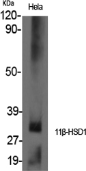

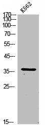
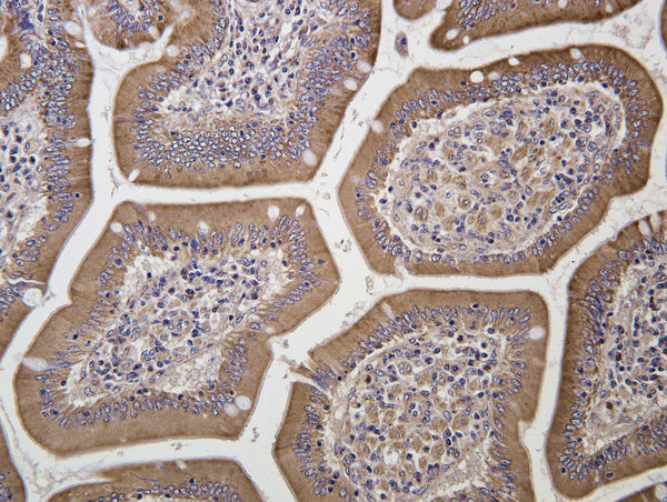
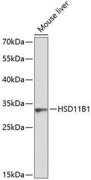
Anti-HSD11B1 Antibody
A95458
ApplicationsWestern Blot, ELISA, ImmunoHistoChemistry
Product group Antibodies
ReactivityHuman, Mouse, Rat
Overview
- SupplierAntibodies.com
- Product NameAnti-HSD11B1 Antibody
- Delivery Days Customer7
- ApplicationsWestern Blot, ELISA, ImmunoHistoChemistry
- CertificationResearch Use Only
- ClonalityPolyclonal
- ConjugateUnconjugated
- HostRabbit
- IsotypeIgG
- Scientific DescriptionRabbit polyclonal antibody to HSD11B1.
- ReactivityHuman, Mouse, Rat
- UNSPSC12352203
Related products
Product group Antibodies
HSD11B1 AntibodyCSB-PA006123
ApplicationsWestern Blot, ELISA
ReactivityHuman, Mouse, Rat
TargetHSD11B1
- SizePrice

Product group Antibodies
Anti-HSD11B1 Antibody Picoband(r)A01565-CARRIER-FREE
ApplicationsWestern Blot, ELISA
ReactivityHuman
TargetHSD11B1
- SizePrice
Product group Antibodies

HSD11B1 / HSD11B AntibodyLS-C831766
ApplicationsELISA, ImmunoHistoChemistry
ReactivityHuman
TargetHSD11B1
- SizePrice
Product group Antibodies

Goat anti-HSD11B1 / HDLEB06597
ApplicationsWestern Blot, ELISA
ReactivityBovine, Human
TargetHSD11B1
- SizePrice

Product group Antibodies
Anti-HSD11B1-25ulHPA047729
ApplicationsWestern Blot, ImmunoHistoChemistry
ReactivityHuman
- SizePrice
Product group Antibodies
HSD11B1 Polyclonal AntibodyCAC13769
ApplicationsImmunoFluorescence, Western Blot, ELISA, ImmunoHistoChemistry
TargetHSD11B1
- SizePrice
Product group Antibodies
References
HSD11B1 Polyclonal AntibodyBS-3904R
ApplicationsImmunoFluorescence, ELISA, ImmunoCytoChemistry, ImmunoHistoChemistry, ImmunoHistoChemistry Frozen, ImmunoHistoChemistry Paraffin
ReactivityBovine, Canine, Equine, Human, Mouse, Porcine, Rabbit, Rat, Sheep
TargetHSD11B1
- SizePrice
Product group Antibodies
HSD11B1 antibodyGTX66405
ApplicationsWestern Blot
ReactivityHuman, Mouse
TargetHSD11B1
- SizePrice