


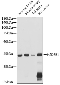
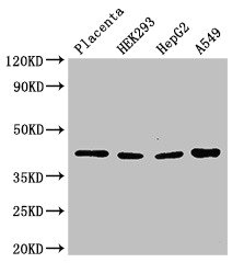
Figure 1. Western blot analysis of HSD3B1 using anti-HSD3B1 antibody (A02856-2). Electrophoresis was performed on a 5-20% SDS-PAGE gel at 70V (Stacking gel) / 90V (Resolving gel) for 2-3 hours. The sample well of each lane was loaded with 50ug of sample under reducing conditions. Lane 1: human placenta tissue lysates, Lane 2: human Caco-2 whole cell lysates, Lane 3: human MDA-MB-453 whole cell lysates. After Electrophoresis, proteins were transferred to a Nitrocellulose membrane at 150mA for 50-90 minutes. Blocked the membrane with 5% Non-fat Milk/ TBS for 1.5 hour at RT. The membrane was incubated with rabbit anti-HSD3B1 antigen affinity purified polyclonal antibody (Catalog # A02856-2) at 0.25 microg/mL overnight at 4°C, then washed with TBS-0.1%Tween 3 times with 5 minutes each and probed with a goat anti-rabbit IgG-HRP secondary antibody at a dilution of 1:10000 for 1.5 hour at RT. The signal is developed using an Enhanced Chemiluminescent detection (ECL) kit (Catalog # EK1002) with Tanon 5200 system. A specific band was detected for HSD3B1 at approximately 42KD. The expected band size for HSD3B1 is at 42KD.
. HSD3B1 was detected in paraffin-embedded section of human placenta tissue. Heat mediated antigen retrieval was performed in EDTA buffer (pH8.0, epitope retrieval solution). The tissue section was blocked with 10% goat serum. The tissue section was then incubated with 1microg/ml rabbit anti-HSD3B1 Antibody (A02856-2) overnight at 4°C. Biotinylated goat anti-rabbit IgG was used as secondary antibody and incubated for 30 minutes at 37°C. The tissue section was developed using Strepavidin-Biotin-Complex (SABC) (Catalog # SA1022) with DAB as the chromogen.")
. Overlay histogram showing CACO-2 cells stained with A02856-2 (Blue line). To facilitate intracellular staining, cells were fixed with 4% paraformaldehyde and permeabilized with permeabilization buffer. The cells were blocked with 10% normal goat serum. And then incubated with rabbit anti-HSD3B1 Antibody (A02856-2, 1microg/1x106 cells) for 30 min at 20°C. DyLight®488 conjugated goat anti-rabbit IgG (BA1127, 5-10microg/1x106 cells) was used as secondary antibody for 30 minutes at 20°C. Isotype control antibody (Green line) was rabbit IgG (1microg/1x106) used under the same conditions. Unlabelled sample without incubation with primary antibody and secondary antibody (Red line) was used as a blank control.")
. HSD3B1 was detected in immunocytochemical section of U20S cells. Enzyme antigen retrieval was performed using IHC enzyme antigen retrieval reagent (AR0022) for 15 mins. The cells were blocked with 10% goat serum. And then incubated with 2microg/mL rabbit anti-HSD3B1 Antibody (A02856-2) overnight at 4°C. DyLight®488 Conjugated Goat Anti-Rabbit IgG (BA1127) was used as secondary antibody at 1:100 dilution and incubated for 30 minutes at 37°C. The section was counterstained with DAPI. Visualize using a fluorescence microscope and filter sets appropriate for the label used.")
Figure 1. Western blot analysis of HSD3B1 using anti-HSD3B1 antibody (A02856-2). Electrophoresis was performed on a 5-20% SDS-PAGE gel at 70V (Stacking gel) / 90V (Resolving gel) for 2-3 hours. The sample well of each lane was loaded with 50ug of sample under reducing conditions. Lane 1: human placenta tissue lysates, Lane 2: human Caco-2 whole cell lysates, Lane 3: human MDA-MB-453 whole cell lysates. After Electrophoresis, proteins were transferred to a Nitrocellulose membrane at 150mA for 50-90 minutes. Blocked the membrane with 5% Non-fat Milk/ TBS for 1.5 hour at RT. The membrane was incubated with rabbit anti-HSD3B1 antigen affinity purified polyclonal antibody (Catalog # A02856-2) at 0.25 microg/mL overnight at 4°C, then washed with TBS-0.1%Tween 3 times with 5 minutes each and probed with a goat anti-rabbit IgG-HRP secondary antibody at a dilution of 1:10000 for 1.5 hour at RT. The signal is developed using an Enhanced Chemiluminescent detection (ECL) kit (Catalog # EK1002) with Tanon 5200 system. A specific band was detected for HSD3B1 at approximately 42KD. The expected band size for HSD3B1 is at 42KD.
Anti-HSD3B1 Antibody Picoband(r)
A02856-2-CARRIER-FREE
ApplicationsFlow Cytometry, ImmunoFluorescence, Western Blot, ELISA, ImmunoCytoChemistry, ImmunoHistoChemistry
Product group Antibodies
ReactivityHuman
TargetHSD3B1
Overview
- SupplierBoster Bio
- Product NameAnti-HSD3B1 Antibody Picoband(r)
- Delivery Days Customer9
- ApplicationsFlow Cytometry, ImmunoFluorescence, Western Blot, ELISA, ImmunoCytoChemistry, ImmunoHistoChemistry
- CertificationResearch Use Only
- ClonalityPolyclonal
- Concentration500 ug/ml
- Gene ID3283
- Target nameHSD3B1
- Target descriptionhydroxy-delta-5-steroid dehydrogenase, 3 beta- and steroid delta-isomerase 1
- Target synonyms3BETAHSD, HSD3B, HSDB3, HSDB3A, SDR11E1, 3 beta-hydroxysteroid dehydrogenase/Delta 5-->4-isomerase type 1, 3 beta-hydroxysteroid dehydrogenase/Delta 5-->4-isomerase type I, 3-beta-HSD I, 3-beta-hydroxy-5-ene steroid dehydrogenase, 3-beta-hydroxy-Delta(5)-steroid dehydrogenase, 3-beta-hydroxysteroid 3-dehydrogenase, delta-5-3-ketosteroid isomerase, dihydrotestosterone oxidoreductase, progesterone reductase, short chain dehydrogenase/reductase family 11E, member 1, steroid Delta-isomerase, trophoblast antigen FDO161G
- HostRabbit
- IsotypeIgG
- Scientific DescriptionBoster Bio Anti-HSD3B1 Antibody Picoband® catalog # A02856-2. Tested in ELISA, Flow Cytometry, IF, IHC, ICC, WB applications. This antibody reacts with Human. The brand Picoband indicates this is a premium antibody that guarantees superior quality, high affinity, and strong signals with minimal background in Western blot applications. Only our best-performing antibodies are designated as Picoband, ensuring unmatched performance.
- ReactivityHuman
- Storage Instruction-20°C,2°C to 8°C
- UNSPSC12352203
Related products
Product group Antibodies
Anti-HSD3B1 AntibodyA16048
ApplicationsImmunoFluorescence, Western Blot, ImmunoCytoChemistry
ReactivityHuman, Mouse, Rat
- SizePrice
Product group Antibodies
Anti-HSD3B1 Antibody144-66533
ApplicationsWestern Blot
ReactivityHuman
TargetHSD3B1
- SizePrice
Product group Antibodies
HSD3B1 Recombinant AntibodyBSM-62395R
ApplicationsWestern Blot
ReactivityHuman
TargetHSD3B1
- SizePrice
Product group Antibodies
HSD3B1 AntibodyCSB-PA010781LA01HU
ApplicationsWestern Blot, ELISA, ImmunoHistoChemistry
ReactivityHuman
TargetHSD3B1
- SizePrice
Product group Antibodies

Goat anti-HSD3B1EB07218
ApplicationsWestern Blot, ELISA, ImmunoHistoChemistry
ReactivityHuman
TargetHSD3B1
- SizePrice
Product group Antibodies
Hsd3B1 Polyclonal AntibodyCAC07863
ApplicationsWestern Blot, ELISA, ImmunoHistoChemistry
TargetHSD3B1
- SizePrice
Product group Antibodies

HSD3B1 AntibodyLS-C401757
ApplicationsELISA, ImmunoHistoChemistry
ReactivityHuman
TargetHSD3B1
- SizePrice

Product group Antibodies
Anti-HSD3B1 AntibodyHPA043261
ApplicationsImmunoHistoChemistry
ReactivityHuman
TargetHSD3B1
- SizePrice
![Various whole cell extracts (30 μg) were separated by 10% SDS-PAGE, and the membranes were blotted with HSD3B1 antibody [N1C1] (GTX114081) diluted at 1:500 and competitor's antibody (sc-30820) diluted at 1:100. The HRP-conjugated anti-rabbit IgG antibody (GTX213110-01) was used to detect the primary antibody.](https://www.genetex.com/upload/website/prouct_img/normal/GTX114081/GTX114081_40436_20170804_WB_competitor_watermark_w_23060501_794.webp)
Product group Antibodies
HSD3B1 antibody [N1C1]GTX114081
ApplicationsWestern Blot, ImmunoHistoChemistry, ImmunoHistoChemistry Paraffin
ReactivityHuman
TargetHSD3B1
- SizePrice